3.2.1.14: chitinase
This is an abbreviated version!
For detailed information about chitinase, go to the full flat file.
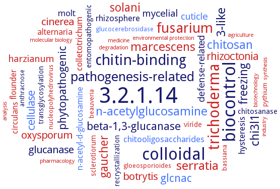
Word Map on EC 3.2.1.14
-
3.2.1.14
-
biocontrol
-
colloidal
-
trichoderma
-
chitin-binding
-
fusarium
-
pathogenesis-related
-
serratia
-
n-acetylglucosamine
-
marcescens
-
chitosan
-
3-like
-
chi3l1
-
solani
-
freezing
-
gaucher
-
glcnac
-
beta-1,3-glucanase
-
phytopathogenic
-
cellulase
-
rhizoctonia
-
oxysporum
-
cinerea
-
botrytis
-
glucanase
-
harzianum
-
mycelial
-
cuticle
-
defense-related
-
hysteresis
-
rhizosphere
-
colletotrichum
-
chitooligosaccharides
-
molt
-
n-acetyl-d-glucosamine
-
alternaria
-
flounder
-
circulans
-
chitosanase
-
entomopathogenic
-
recrystallization
-
sclerotiorum
-
transglycosylation
-
viride
-
beauveria
-
bassiana
-
pythium
-
nucleopolyhedrovirus
-
anthracnose
-
glucocerebrosidase
-
gloeosporioides
-
environmental protection
-
molecular biology
-
analysis
-
synthesis
-
pharmacology
-
industry
-
degradation
-
medicine
-
biotechnology
-
agriculture
- 3.2.1.14
-
biocontrol
-
colloidal
- trichoderma
-
chitin-binding
- fusarium
-
pathogenesis-related
- serratia
- n-acetylglucosamine
- marcescens
- chitosan
-
3-like
-
chi3l1
- solani
-
freezing
- gaucher
- glcnac
- beta-1,3-glucanase
-
phytopathogenic
- cellulase
- rhizoctonia
- oxysporum
- cinerea
- botrytis
- glucanase
- harzianum
- mycelial
- cuticle
-
defense-related
-
hysteresis
-
rhizosphere
- colletotrichum
- chitooligosaccharides
-
molt
- n-acetyl-d-glucosamine
- alternaria
- flounder
- circulans
- chitosanase
-
entomopathogenic
-
recrystallization
- sclerotiorum
-
transglycosylation
- viride
- beauveria
- bassiana
- pythium
- nucleopolyhedrovirus
-
anthracnose
- glucocerebrosidase
- gloeosporioides
- environmental protection
- molecular biology
- analysis
- synthesis
- pharmacology
- industry
- degradation
- medicine
- biotechnology
- agriculture
Reaction
Synonyms
1,4-beta-poly-N-acetylglucosaminidase, AcD1ChiA, acid mammalian chitinase, acidic chitinase, acidic mammalian chitinase, active phase-associated protein I, active phase-associated protein II, AD2PF-ChiA, AgChi, alpha-chitinase, AMCase, antifreeze protein, APAP1, APAP2, APAPI, APAPII, AtChiC, B220, bacterial-type chitinase, Bbchit1, BChi14, beta-1,4-poly-N-acetyl glucosamidinase, BjCHI1, BJL200-ChiC1, CF-AG, CF-antigen, CGCHI3, CH5B, ChBDChiC, CHI, Chi-1, CHI-26, Chi-A, Chi-h, Chi1, Chi18A, Chi18B, Chi18C, Chi19, CHI2, Chi2 enzyme, Chi21, Chi22, Chi3, Chi31, Chi35, Chi40, Chi44, Chi46, Chi54, CHI60, Chi67, Chi70, Chi72, Chi78, ChiA, ChiA Nima, ChiA protein, ChiA-HD73, ChiA-Hh59, ChiA-Mt45, ChiA1, ChiA74, ChiB, ChiC, ChiCW, ChiD, ChiE, ChiEN1, ChiF, ChiF1, ChiG, ChiNCTU2, Chit, CHIT 1A, CHIT 1B, CHIT1, Chit1b, Chit33, CHIT42, Chit62, ChitA, ChitIII-1, ChitIII-2, chitinase, chitinase 1, chitinase 18C, chitinase 2, chitinase 60, chitinase 92, chitinase A, chitinase A1, chitinase B, chitinase C, chitinase C1, chitinase Chi255, chitinase F1, chitinase G, chitinase I, chitinase II, chitinase III, chitinase-A, chitinases A, chitinolytic enzyme, chitodextrinase-N-, chitotriosidase, ChiW, CHT1, Cht4, CHT7, ChtII, ChtIII, CiX1, class I chitinase, class II chitinase, class III chitinase, class IV chitinase, class V chitinase, Complement-fixation antigen, CpCHI, CrChi1, CTS1, ECH, ECH1, Ech42, ech46, ELC, endo-chitinase, endochitinase, endochitinase 1, endochitinase-1, endochitinase-2, exochitinase, GAC1, GAC2, GH18 chitinase, group I chitinase, group II chitinase, group III chitinase, hevamine, HoChiA, HoChiB, HoChiC, HschiA1, Is-chiB, LlChi18A, LpChiA, MACase, MF1 antigen, MmChi60, More, MsChi386, MsChi535, NaCHIT1, NaCHIT3, NtChitIV, NtChiV, OfChtI, OsChia1b, OsChia1c, OsChia1cDELTAChBD, OsChia1d, OsChia2a, OsChia2b, OsChia4a, OsChib1a, PF1233, PjChi-1, plant class III chitinase, PLC-A, PLC-B, PLChiA, poly-beta-1, 4-[2-acetamido-2-deoxy]-delta-glucoside glycanohydrolase, poly-beta-glucosaminidase, PrChi-A, Ptchi19p, Ra-ChiC, RCB4, RHBC, sAMC, SCChi-1, SI-CLP, SjChi, SsChi50, stabilin-1 interacting chitinase-like protein, STK_09390, TBC-1, Tfu_0580, Tk-ChiA, TKU028 chitinase, TmChi, TSA1902, UDA, V-CHIA, VpChiA, WIN6, YlCts1p, Ym1
ECTree

Advanced search results
Crystallization
Crystallization on EC 3.2.1.14 - chitinase
Please wait a moment until all data is loaded. This message will disappear when all data is loaded.
 top
topCRYSTALLIZATION (Commentary) 

ORGANISM
UNIPROT
LITERATURE
purified recombinant AtChiC, sitting drop vapor diffusion method, mixing of 0.001 ml of 5 mg/ml protein in water with 0.001 ml of reservoir solution containing 100 mM trisodium citrate, pH 5.6, and 20% PEG 10000, 20°C, 5 months, X-ray diffraction structure determination and analysis at 2.0 A resolution, molecular replacement
Q0WT03
native enzyme with precipitant PEG 3350, at 18°C, X-ray diffraction structure determination and analysis at 1.7-2.1 A resolution
-
hanging-drop vapour-diffusion method. According to the diffraction of chitinase crystals at 1.10 A resolution, the crystal belongs to space group P2(1), with unit-cell parameters a = 50.79, b = 48.79, c = 66.87 A, beta = 99.31°. There is one chitinase molecule in the asymmetric unit, with a solvent content of 43.4%
-
sitting-drop vapor diffusion at room temperature. X-ray structures of the chitinase catalytic domain from wild-type (apo, as well as with chloride ions bound) and a E234A mutant enzyme, solved by molecular replacement and refined at 1.53, 1.8 and 1.7 A resolution, respectively
-
purified native enzyme, hanging drop vapor diffusion method, 0.17 M Li2SO4, 0.085 M Tris-HCl, pH 8.5, 25.5% PEG 4000, 15% glycerol, and in the presence of a large excess of GlcNAc, binding to (GlcNAc)4, X-ray diffraction structure determination and analysis at 1.5 A resolution, molecular replacement
purified enzyme CrChi1 free or in complex with inhibitor caffeine, hanging drop vapour diffusion method, 0.0005 ml protein solution, containing 20 mg/ml in 20 mM Tris/HCl, pH 8.0, and 0.0005 ml reservoir solution are mixed and equilibrated against 0.2 ml reservoir solution consisting of 0.2 M ammonium dihydrogen phosphate, and 15% w/v PEG3350, for the enzyme complex addition of 0.001 ml of 1 mM caffeine solution, 16°C, X-ray diffraction structure determination and analysis at 1.8 A and 1.6 A, respectively, molecular replacement
purified recombinant His-tagged CrChi1, hanging drop vapour diffusion method, 0.0005 ml of 20 mg/ml protein in 20 mM Tris-HCl buffer pH 8.0 is mixed with an equal volume of precipitant solution containing containing 0.2 M ammonium dihydrogen phosphate and 20% w/v PEG 3350, pH 4.6., equilibration against 0.2 ml of reservoir solution at 18°C, X-ray diffraction structure determination and analysis at 1.8 A resolution
-
purified enzyme, hanging drop vapour diffusion method, 0.002 m of 16 mg/ml protein in 20 mM acetate, pH 5.0, is mixed with 0.002 ml of reservoir solution containing PEG 8000, equilibration over 1.0 ml reservoir solution, 20°C, 3 days, X-ray diffraction structure determination and analysis at 2.1 A resolution, molecular replacement
-
AMCase catalytic domain in the apo form and in complex with the inhibitor methylallosamidin, X-ray diffraction structure determination and analysis at 1.7-2.0 A resolution
purified recombinant enzyme in complex with inhibitor 1, hanging drop vapor diffusion, 0.0002 ml of protein solution containing 0.44 mM AMCase, 0.52 mM inhibitor 1, 25 mM Tris-HCl, pH 8.0, and 50 mM NaCl, is mixed with 0.0002 ml of reservoir solution containing 20% PEG MME 5000, 100 mM bis-Tris, equilibration over 0.2 ml of reservoir solution, pH 6.5, 18°C, 2 weeks, X-ray diffraction structure determination and analysis, modeling
-
three-dimensional modeling of variant isoforms. The overall protein fold is not altered in mutant N45D/D47N/R61M
purified recombinant native and SeMet-labeled wild-type enzymes and mutant E115Q enzyme, hanging drop vapor diffusion method, mixing of 0.001 ml 5 mg/ml protein in water with 0.001 ml of reservoir solution containing 100 mM Tris-HCl, pH 8.5, and 1.7-2.2 M ammonium dihydrogen phosphate, 20°C, 1 months, X-ray diffraction structure determination and analysis at 1.2, 1.6, and 1.8 A resolution, respectively, single-wavelength anomalous dispersion method
recombinant enzyme from 18% w/v PEG 8000, 0.2 M zinc acetate dehydrate, and 0.1 M sodium cacodylate, pH 6.5, X-ray diffraction structure determination and analysis at 1.2 A resolution, multiwavelength anomalous diffraction method with zinc as the anomalous scatter
-
catalytically active domains ChtII-C1 and ChtII-C2, both in unliganded form and complexed with chitooligosaccharide substrates, hanging drop vapor diffusion method, using 0.2 M sodium chloride, 0.1 M Tris, pH 8.5, and 25% (w/v) PEG3350 for ChtII-C1 and 0.2 M ammonium acetate, 0.1 M Bis-Tris, pH 6.5, and 45% (w/v) 2-methyl-2,4-pentadiol for ChtII-C2
hanging drop vapor diffusion method, using 200 mM ammonium sulfate, 100 mM bis-Tris pH 6.5, 20% (w/v) PEG 3350 for domain GH18A, and 200 mM trisodium citrate dihydrate pH 8.1, 20% (w/v) PEG 3350 for domain GH18B
purified recombinant His6-tagged wild-type enzyme and mutant E148Q unliganded, and wild-type enzyme and mutant E148A in complex with chitobiose and /or chitotriose, hanging drop vapour diffusion method, mixing of 0.001 ml of 10 mg/ml protein in 50 mM HEPES, pH 8.0, and 100 mM NaCl, with 0,001 ml of reservoir solution containing 100 mM HEPES, pH 7.5, 25% w/v PEG 3350 for the wild-type with soaking of crystals in ligand containing solution for the complex crystals, reservori solution containing 5 mM (GlcNAc)6 in 100 mM HEPES, pH 7.9, 23% w/v PEG 3350 and in 100 mM HEPES, pH 8.0, 21% w/v PEG 3350, respectively, for mutant E148A and E148Q, X-ray diffraction structure determination and analysis at 1.7 A and 2.2 A resolution
catalytic domain, in complex with beta-D-N,N',N''-triacetylchitotriose or beta-D-N,N'-diacetylchitobiose, sitting drop vapor diffusion method, using 0.8-1.3 M (NH4)2HPO4 and 0.1 M sodium citrate buffer, pH 4.5-5.5
homology modeling using the human chitotriosidase structure as template, PDB 1LG1
bi-lobed structure with a wide cleft lined by conserved residues. Glu113 acts as the proton donor, and Glu122 as a general base
catalytic domain of chitinase recombinantly expressed in Escherichia coli, hanging-drop vapour diffusion method, resolution of 1.50 A. The crystals belong to space group P2(1)2(1)2(1), with unit-cell parameters a = 90.0, b = 92.8, c = 107.2 A
hanging drop vapor diffusion method, crystallographic analyses of the catalytic domain of chitinase. Crystals of these mutant enzymes E526A and D524A in complex with chito-oligosaccharide substrate by means of cocrystallization and soaking methods, respectively, and determination of their tertiary structures. X-ray diffraction data are collected, resolutions of 1.80 A and 1.76 A
the crystal structure of the catalytic domain of a chitinase is determined at 1.5 A resolution
-
wild-type and mutant ChBD2 domains, X-ray diffraction structure determination and analysis at 1.7-1.8 A resolution
-
purified isolated interdomain loop Ra-ChiC89-252, hanging drop vapour diffusion method, mixing of 0.002 ml of protein solution with 0.002 ml of reservoir solution at 20°C, equilibration against reservoir solution, X-ray diffraction structure determination and analysis at 1.85-1.90 A resolution, presence of three, four or five Ra-ChiC89-252 molecules in the asymmetric unit, respectively
-
purified wild-type enzyme and mutants E141Q and E162Q, hanging-drop vapor diffusion method, mixing of 4.5-6.5 mg/ml protein with reservoir solution, crystals of E141Q and E162Q mutants are obtained by the micro-seeding method using wild-type crystals as a seed, ligand-bound forms are obtained by the soaking method, 20°C, X-ray diffraction structure determination and analysis at 1.9-2.15 A resolution
crystallization of the native enzyme and the enzyme in complex with 8-chlorotheophylline, kinetin or acetazolamide, sitting drop method, 1.6 A resolution
crystal structure analysis of ChiB bound to argadin or to argifin, molecular modelling, overview
hanging-drop, vapor-diffusion method at 20°C, crystal structure of full-length ChiC, wild-type enzyme and mutant enzyme E147Q at 2.45 A and 2.0 A resolution
-
14 mg/ml purified chitinase in 100 mM Tris, pH 7.4, sitting drop vapour diffusion method, mixing of 0.002 ml protein solution with the same volume of reservoir solution containing 100 mM MES pH 6.0, 10% w/v PEG 4000, at 20°C, X-ray diffraction structure determination and analysis at 2.6 A resolution
-
purified recombinant His6-tagged chitinase A 575 amino acid fragment, from a solution containing 10% PEG 400, 0.1 M sodium acetate, pH 4.6, and 0.125 M CaCl2, X-ray diffraction structure determination and analysis at 2.14 A resolution
-
purified recombinant His6-tagged wild-type and mutant E315M mutant enzymes, free or bound to (GlcNAC)5 or (GlcNAc)6, sitting drop vapour diffusion method, 20 nl of protein solution containing 20 mg/ml wild-type protein in 10 mM Tris-HCl, pH 8.0, are mixed with 20 nl of precipitant solution, macroseeding, or 5 mg/ml protein in 1.2 M ammonium sulfate and 0.1 M Tris-HC, pH 8.0, at 14°C for 2 days, mutant E315M crystals from 20% w/v PEG 4000, 0.1 M ammonium sulfate and 0.1 M Tris-HCl, pH 7.5, 1 week at 14°C, X-ray diffraction structure determination and analysis at 1.7-2.0 A resolution
-