3.5.1.1: asparaginase
This is an abbreviated version!
For detailed information about asparaginase, go to the full flat file.
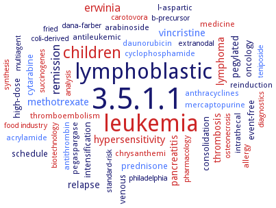
Word Map on EC 3.5.1.1
-
3.5.1.1
-
leukemia
-
lymphoblastic
-
children
-
remission
-
erwinia
-
hypersensitivity
-
methotrexate
-
lymphoma
-
vincristine
-
pegylated
-
relapse
-
pancreatitis
-
thrombosis
-
prednisone
-
high-dose
-
venous
-
oncology
-
event-free
-
schedule
-
cytarabine
-
consolidation
-
intensification
-
antileukemic
-
intrathecal
-
antithrombin
-
l-aspartic
-
chrysanthemi
-
anthracyclines
-
medicine
-
reinduction
-
cyclophosphamide
-
pegaspargase
-
thromboembolism
-
allergy
-
acrylamide
-
arabinoside
-
mercaptopurine
-
daunorubicin
-
osteonecrosis
-
analysis
-
philadelphia
-
extranodal
-
fried
-
b-precursor
-
succinogenes
-
standard-risk
-
synthesis
-
coli-derived
-
food industry
-
diagnostics
-
biotechnology
-
dana-farber
-
pharmacology
-
teniposide
-
carotovora
-
multiagent
- 3.5.1.1
- leukemia
- lymphoblastic
- children
-
remission
- erwinia
- hypersensitivity
- methotrexate
- lymphoma
- vincristine
-
pegylated
-
relapse
- pancreatitis
- thrombosis
- prednisone
-
high-dose
- venous
-
oncology
-
event-free
-
schedule
- cytarabine
-
consolidation
-
intensification
-
antileukemic
-
intrathecal
- antithrombin
-
l-aspartic
- chrysanthemi
- anthracyclines
- medicine
-
reinduction
- cyclophosphamide
-
pegaspargase
- thromboembolism
- allergy
- acrylamide
-
arabinoside
- mercaptopurine
- daunorubicin
- osteonecrosis
- analysis
-
philadelphia
-
extranodal
-
fried
-
b-precursor
- succinogenes
-
standard-risk
- synthesis
-
coli-derived
- food industry
- diagnostics
- biotechnology
-
dana-farber
- pharmacology
- teniposide
- carotovora
-
multiagent
Reaction
Synonyms
14270 ASNase, alpha-asparaginase, AnsA, ansA3, AnsB, ansZ, Asn, ASNase, ASNase1, ASNase3, ASP1, ASP3, asparaginase, asparaginase II, asparagine amidohydrolase, asparginase II, ASPG, ASPG II, ASPGA1, ASPGB1, ASRGL1, At3g16150, colaspase, crasnitin, DiAsp, EcAII, EcAIII, EcaL-ASNase, ECAR-LANS, elspar, ErA, Erwinase, glutaminase-free L-asparaginase, glutamine-(asparagin-)ase, HPA, L-ASNase, L-asparaginase, L-asparaginase 2, L-asparaginase I, L-asparaginase II, L-asparaginase III, L-asparaginase type II, L-asparaginase type III, L-asparaginase-I, L-asparaginase-II, L-asparagine amidohydrolase, L-asparagine amino hydrolase, L-asparagine aminohydrolase, leunase, PfA, PhA, potassium-dependent asparaginase, potassium-independent asparaginase, potassium-independent L-asparaginase, SAMN05216263_10744, SGR ASNase, type I L-asparaginase, type II L-asparaginase, Ypa
ECTree

Advanced search results
Crystallization
Crystallization on EC 3.5.1.1 - asparaginase
Please wait a moment until all data is loaded. This message will disappear when all data is loaded.
 top
topCRYSTALLIZATION (Commentary) 

ORGANISM
UNIPROT
LITERATURE
hanging drop vapor diffusion method, using 0.2 M ammonium sulfate, 0.1 M MES at pH 6.0, and 13-15% (w/v) PEG 3350
-
sitting drop vapor diffusion method, using 0.1 M HEPES, pH 7.0, 15% (w/v) PEG 4000
complexed with the L- and D-stereoisomers of the suicide inhibitor L-6-diazo-5-oxy-norleucine and D-6-diazo-5-oxynorleucine solved using X-ray crystallography and refined with data extending to 1.7 A
crystal structures in complex with L-aspartic acid and with L-glutamic acid. The enzyme conformations open and closed correspond to the inactive and active states, respectively. The binding of ligands induces the positioning of the catalytic Thr15 into its active conformation, which in turn allows for the ordering and closure of the flexible N-terminal loop. L-Aspartic acid is more efficient than L-glutamic acid in inducing the active positioning of Thr15
high-resultion crystal structures of the complex of L-asparaginase with L-Glu, D-Asp and succinic acid
hanging-drop vapour-diffusion method, X-ray structure of the enzyme, crystallized in a new form, space group C2 and refined to 1.95 A resolution, is compared with that of the previously determined crystal for, space group P2(1)
-
mutant enzyme D90E, hanging drop vapor diffusion method, using 25% PEG MME 550, 100 mM MES pH 6.5, 10 mM ZnSO4 or 30% (w/v) PEG MME 550, 100 mM bicine pH 9.0, 100 mM NaCl or 100 mM HEPES pH 7.5, saturated solution of tribasic sodium citrate mixed with buffer, at citrate:buffer ratio of 9:1
mutant enzyme Y25F, untreated crystals and crystals soaked with L-hydroxylysine. Comparison with previously reported structures. The loop acting as a gate over the active site is very flexible. Its structure in the native enzyme is primarily controlled by the occupancy of the active site
the aspartate product in the crystal structure of L-ASP exists in an unusual alpha-COOH protonation state. The crystal structures may represent intermediate steps rather than initial binding. The substrate's alpha-carboxyl may serve as a proton acceptor and activate one of the catalytic threonines during L-ASP's nucleophilic attack on the amide carbon
resolution of 1.4 A. There are major differences in the active site flexible loop and in the 286-297 loop from the second subunit, which is involved in active-site formation. Accordingly, Glu289, Asn255, and Gln63 are suggested to play roles in modulating the accessibility of the active site
hanging drop vapor diffusion method, using 2.2-2.5 M sodium malonate (pH 7.0)
purified recombinant enzyme, protein solution at 10 mg/ml is mixed in 2:1 ratio with a solution containing 25% w/v PEG 4000, 100 mM HEPES, pH 6.5, and 200 mM MgCl2, incubation overnight at room temperature, the precipitate is centrifuged and the supernatant is diluted 1:1 with water, was used for hanging-drop vapor diffusion, 0.003 ml solution is mixed with 0.001 ml of reservoir solution containing 20% PEG 4000, 100 mM HEPES, pH 6.5, and 200 mM MgCl2, 200 mM MgCl2, streak-seeding with nuclei, room temperature, one day, X-ray diffraction structure determination and analysis at 2.6 A resolution, structure modelling
-
crystals are grown by the hanging-drop vapour-diffusion technique at 22°C. The crystal structures of Erwinia carotovora L-asparaginase complexed with L-aspartate and L-glutamate are determined at 1.9 A and 2.2 A, respectively
-
purified recombinant L-asparaginase in the presence of L-glutamate, hanging drop vapour diffusion method, 10 mg ml protein with 10 mM glutamate, in 1618% w/v PEG 3350, 10 mM phosphate, pH 7.0, and 0.2 M NaF, 20°C, X-ray diffraction structure determination and analysis at 2.6 A resolution using an in-house rotating-anode generator, crystals of monoclinic P21 space group, molecular-replacement
-
purified recombinant enzyme, hanging drop vapour diffusion method, 0.002 ml or protein solution is mixed with 0.001 ml reservoir solution containing 0.1 M Tris-HCl, pH 8.5, 12% v/v PEG 400, and 30 mM NaCl, soaking of crystals in 25% v/v PEG 400 and flash-cooling, X-ray difraction structure determination and analysis at 2.16 A resolution
-
wild-type and mutant S121P. Residue 121 impacts the conformation of the conserved tyrosine 27, a component of the catalytically-important flexible N-terminal loop