1.1.1.40: malate dehydrogenase (oxaloacetate-decarboxylating) (NADP+)
This is an abbreviated version!
For detailed information about malate dehydrogenase (oxaloacetate-decarboxylating) (NADP+), go to the full flat file.
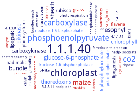
Word Map on EC 1.1.1.40
-
1.1.1.40
-
chloroplast
-
co2
-
carboxylase
-
phosphoenolpyruvate
-
maize
-
bundle
-
sheath
-
glucose-6-phosphate
-
mesophyll
-
carboxykinase
-
thioredoxins
-
dikinase
-
chlorophyll
-
grass
-
rubisco
-
photosystems
-
sorghum
-
nad-malic
-
6-phosphogluconate
-
lipogenesis
-
lipogenic
-
flaveria
-
fructose-1,6-bisphosphatase
-
orthophosphate
-
ribulose-1,5-bisphosphate
-
kranz
-
nadp-isocitrate
-
pck
-
crassulacean
-
photorespiration
-
nadph-generating
-
4.1.3.8
-
calvin
-
trinervia
-
phosphoribulokinase
-
non-photosynthetic
-
ferredoxin-thioredoxin
-
c4-like
-
bundle-sheath
-
photorespiratory
-
6.4.1.2
-
bidentis
-
crystallinum
-
3.1.3.11
-
4.1.1.31
-
fbpase
-
water-use
-
panicum
-
mesembryanthemum
-
nadp-icdh
-
analysis
-
biotechnology
-
medicine
- 1.1.1.40
- chloroplast
- co2
- carboxylase
- phosphoenolpyruvate
- maize
- bundle
- sheath
- glucose-6-phosphate
- mesophyll
-
carboxykinase
- thioredoxins
- dikinase
- chlorophyll
- grass
- rubisco
-
photosystems
- sorghum
-
nad-malic
- 6-phosphogluconate
-
lipogenesis
-
lipogenic
- flaveria
- fructose-1,6-bisphosphatase
- orthophosphate
- ribulose-1,5-bisphosphate
-
kranz
-
nadp-isocitrate
- pck
-
crassulacean
-
photorespiration
-
nadph-generating
-
4.1.3.8
-
calvin
- trinervia
- phosphoribulokinase
-
non-photosynthetic
-
ferredoxin-thioredoxin
-
c4-like
-
bundle-sheath
-
photorespiratory
-
6.4.1.2
- bidentis
- crystallinum
-
3.1.3.11
-
4.1.1.31
- fbpase
-
water-use
- panicum
- mesembryanthemum
- nadp-icdh
- analysis
- biotechnology
- medicine
Reaction
Synonyms
ADP-malic enzyme2, c-NADP-ME, C4 NADP-malic enzyme, C4 photosynthetic NADP-malic enzyme, C4-NADP-malic enzyme, C4-NADP-ME, ChlME1, ChlME2, cNAD-ME, cytoNADPME, cytosolic malic enzyme, cytosolic NADP+-dependent isoform, cytosolic NADP+-dependent malic enzyme, L-malate: NADP oxidoreductase (decarboxylating), L-malate: NADP oxidoreductase [OAA decarboxylating], L-malate: NADP oxidoreductase [oxaloacetate decarboxylating], L-malate:NADP oxidoreductase, L-malate:NADP oxidoreductase (oxaloacetate decarboxylating), m-NAD(P)-ME, m-NADP-ME, MaeB, MaeB1, MalA, malate dehydrogenase (decarboxylating, NADP), malate dehydrogenase (NADP, decarboxylating), malE1, malic enzyme, malic enzyme 1, malic enzyme 2, malic enzyme 3, malic enzyme-NADP, ME, ME-61, ME-70, ME-NADP, ME1, ME2, ME3, mitochondrial malic enzyme, mitochondrial NADP malic enzyme, mNAD-ME, NAD(P)+-malic enzyme, NADP dependent malic enzyme, NADP malic enzyme, NADP(+)-dependent mitochondrial malic enzyme 2, NADP(H)-dependent malic enzyme, NADP+ dependent malic enzyme, NADP+-dependent decarboxylating malate dehydrogenase, NADP+-dependent malic enzyme, NADP+-dependent malic enzyme 3, NADP+-dependent ME, NADP+-ME, NADP-dependent malate dehydrogenase, NADP-dependent malic enzyme, NADP-dependent malic enzyme 1, NADP-dependent ME, NADP-linked decarboxylating malic enzyme, NADP-malate dehydrogenase, NADP-malate enzyme, NADP-malic enzyme, NADP-malic enzyme 1, NADP-malic enzyme 2, NADP-MDH, NADP-ME, NADP-ME1, NADP-ME2, NADP-ME3, NADP-ME4, NADP-specific malate dehydrogenase, NADP-specific malic enzyme, NADP-specific ME, NADPH-dependent malic enzyme, NADPH-dependent malic enzyme 1, NADPH-dependent ME1, nicotinamide adenine dinucleotide phosphate-dependent malic enzyme, nicotinamide adenine dinucleotide phosphate-malic enzyme, nonC4-NADP-ME, pNAD-ME, pyruvic-malic carboxylase, RHA1_RS44255, TME
ECTree

Advanced search results
Subunits
Subunits on EC 1.1.1.40 - malate dehydrogenase (oxaloacetate-decarboxylating) (NADP+)
 top
top

