3.4.24.18: meprin A
This is an abbreviated version!
For detailed information about meprin A, go to the full flat file.
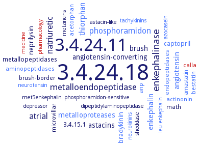
Word Map on EC 3.4.24.18
-
3.4.24.18
-
3.4.24.11
-
enkephalinase
-
metalloendopeptidase
-
atrial
-
phosphoramidon
-
natriuretic
-
enkephalin
-
thiorphan
-
angiotensin-converting
-
angiotensin
-
astacins
-
metalloproteases
-
brush
-
bradykinin
-
3.4.15.1
-
captopril
-
neprilysin
-
metallopeptidases
-
anp
-
aminopeptidases
-
acetorphan
-
math
-
endopeptidases
-
brush-border
-
microvillar
-
actinonin
-
calla
-
bestatin
-
amastatin
-
azocasein
-
phosphoramidon-sensitive
-
sheddase
-
dipeptidylaminopeptidase
-
metzincins
-
pharmacology
-
neurokinins
-
astacin-like
-
met5enkephalin
-
depressor
-
medicine
-
tachykinins
-
neurotensin
-
leu-enkephalin
- 3.4.24.18
-
3.4.24.11
- enkephalinase
- metalloendopeptidase
- atrial
- phosphoramidon
-
natriuretic
- enkephalin
- thiorphan
-
angiotensin-converting
- angiotensin
- astacins
- metalloproteases
-
brush
- bradykinin
-
3.4.15.1
- captopril
- neprilysin
- metallopeptidases
- anp
- aminopeptidases
- acetorphan
-
math
- endopeptidases
-
brush-border
- microvillar
- actinonin
- calla
- bestatin
- amastatin
- azocasein
-
phosphoramidon-sensitive
- sheddase
-
dipeptidylaminopeptidase
-
metzincins
- pharmacology
- neurokinins
-
astacin-like
-
met5enkephalin
-
depressor
- medicine
- tachykinins
- neurotensin
- leu-enkephalin
Reaction
Hydrolysis of protein and peptide substrates preferentially on carboxyl side of hydrophobic residues =
Synonyms
E-24.18, EC 3.4.24.11, Endopeptidase-2, M12.002, MEP1A, Meprin, meprin A, meprin A metalloprotease, meprin A metalloproteinase, meprin A subunit alpha, meprin alpha, meprin alpha1, meprin alpha2, meprin beta, meprin metalloprotease, meprin metalloproteinase, Meprin-a, meprin-alpha, metalloprotease meprin A, metalloproteinase meprin alpha, Mmepa, More, mouse meprin alpha, N-Benzoyl-L-tyrosyl-p-aminobenzoic acid hydrolase, PABA peptide hydrolase, PABA-peptide hydrolase, PPH, PPH alpha, PPH beta, procollagen proteinase, Rmepa
ECTree

Advanced search results
General Information
General Information on EC 3.4.24.18 - meprin A
Please wait a moment until all data is loaded. This message will disappear when all data is loaded.
 top
topGENERAL INFORMATION 

ORGANISM
UNIPROT
COMMENTARY 
LITERATURE
evolution
malfunction
metabolism
physiological function
additional information
evolution

-
meprin A, is a membrane-associated neutral metalloendoprotease that belongs to the astacin family of zinc endopeptidases
evolution
-
meprin A, is a membrane-associated neutral metalloendoprotease that belongs to the astacin family of zinc endopeptidases
evolution
-
meprin A, is a membrane-associated neutral metalloendoprotease that belongs to the astacin family of zinc endopeptidases
evolution
-
meprin alpha is a metalloprotease of the astacin family characterized by a conserved zinc-binding motif (HExxHxxGFxHExxRxDR). Human meprin-alpha and -beta protease, EC 3.4.24.63, subunits are 55% identical at the amino acid level, while the substrate and peptide bond specificities vary markedly
evolution
-
meprin metalloproteases belong to the astacin family of zinc endopeptidases and the metzincin superfamily
evolution
-
meprin metalloproteases belong to the astacin family of zinc endopeptidases and the metzincin superfamily
evolution
-
meprin metalloproteases belong to the astacin family of zinc endopeptidases and the metzincin superfamily. Meprins belong to the astacin family of metalloproteases, comprising only six members in humans. These enzymes are characterized by a conserved zinc-binding motif (HExxHxxGxxHxxxRxDR) and by a sequence in close proximity to the active-site cleft, the so called Met-turn, that includes a tyrosine residue as a fifth zinc ligand. Within the astacin family, meprins exhibit a unique domain composition
evolution
meprin alpha belongs to the astacin family of zinc-endopeptidases and the metzincin superfamily, characterized by the conserved motif HExxHxxGxxHxxxRxDR
evolution
the astacin proteases meprin alpha and meprin beta are zinc-dependent metalloproteases of the metzincin superfamily
evolution
the enzyme encoded by Mmepa belongs to the BTP cluster of the astacin enzyme family. Structure-activity relationship of astacin metalloproteases, EDTA is used to dock into the active site cleft of the astacins to know the interaction network and to identify the important residues for binding, comparative three-dimensional structure homology modeling and docking study, and potential binding site, detailed overview
evolution
the enzyme encoded by Rmepa belongs to the BTP cluster of the astacin enzyme family. Structure-activity relationship of astacin metalloproteases, EDTA is used to dock into the active site cleft of the astacins to know the interaction network and to identify the important residues for binding, comparative three-dimensional structure homology modeling and docking study, and potential binding site, detailed overview
evolution
-
meprin A, is a membrane-associated neutral metalloendoprotease that belongs to the astacin family of zinc endopeptidases
-
malfunction
knockdown of meprin alpah1 mRNA causes defects in general tissue differentiation
malfunction
meprin alpha2 morphants show severe failures in the formation of the vascular system
malfunction
-
meprin inhibition elevates levels of natriuretic peptides in plasma and the vascular wall, decreases plaque volume and suppresses lipid deposition in carotid arteries, reduces production of reactive oxygen species and apoptosis (which are associated with atherosclerosis) in the vascular wall, and increases natriuretic peptide function on cell apoptosis, proliferation, and intracellular reactive oxygen species generation in the THP-1 cell line and primary vascular smooth muscle cells
malfunction
-
altered localization and shedding of meprin A in places other than the apical membranes may be deleterious in vivo in acute tubular injury. Importance of a sheddase involved in the release of membrane-associated meprin A under pathological conditions
malfunction
-
altered localization and shedding of meprin A in places other than the apical membranes may be deleterious in vivo in acute tubular injury. Importance of a sheddase involved in the release of membrane-associated meprin A under pathological conditions
malfunction
-
breast cancer MDA-MB-435 cells treated with the meprin inhibitor actinonin are less invasive in vitro. Altered localization and shedding of meprin A in places other than the apical membranes may be deleterious in vivo in acute tubular injury. Importance of a sheddase involved in the release of membrane-associated meprin A under pathological conditions
malfunction
-
enzyme downregulation causes impaired intestinal mucin release and barrier function,and decreases tensile strength in the skin, but it also leads to protection against sepsis and renal injury. Enzyme upregulation can cause fibrosis, pulmonary hypertension, the Kawasaki syndrome, inflammatory bowel disease, and is involved in nephritis, cancer, and Alzheimer's disease, overview
malfunction
-
enzyme-deficient mice show lower amounts of mature collagen I compared with wild-type mice and exhibit significantly reduced collagen deposition in skin, along with markedly decreased tissue tensile strength
malfunction
-
meprin alpha knock-out mice exhibit decreased collagen deposition in skin resulting in impaired tensile strength, overview. Overexpression of meprin metalloproteases occurs under fibrotic conditions in the skin (keloids) and the lung (pulmonary hypertension)
malfunction
-
mice lacking meprin alpha and meprin beta are significantly protected against renal ischaemia/reperfusion injury and bladder inflammation. Meprin alpha-knockout mice exhibit less renal damage compared with wild-type mice
malfunction
-
monocytes from meprin knockout mice on a C57BL/6 background are less able to migrate through an MDCK cell monolayer than monocytes from their wild-type counterparts
malfunction
-
the knockdown of meprin beta in zebrafish embryos leads to a general failure in organogenesis, resulting in the death of the embryos between days 1 and 3 postfertilization
malfunction
-
monocytes from meprin knockout mice on a C57BL/6 background are less able to migrate through an MDCK cell monolayer than monocytes from their wild-type counterparts
-
malfunction
-
altered localization and shedding of meprin A in places other than the apical membranes may be deleterious in vivo in acute tubular injury. Importance of a sheddase involved in the release of membrane-associated meprin A under pathological conditions
-
metabolism
-
following ischemia-reperfusion- and cisplatin-induced acute kidney injury, meprin A is redistributed toward the basolateral plasma membrane, and the cleaved form of meprin A is excreted in the urine
metabolism
-
following ischemia-reperfusion- and cisplatin-induced acute kidney injury, meprin A is redistributed toward the basolateral plasma membrane, and the cleaved form of meprin A is excreted in the urine
metabolism
-
following ischemia-reperfusion- and cisplatin-induced acute kidney injury, meprin A is redistributed toward the basolateral plasma membrane, and the cleaved form of meprin A is excreted in the urine
metabolism
-
meprin A is more effective than meprin B, EC 3.4.24.63, in impairing MDCK epithelial barrier function
metabolism
-
meprins show higher substrate and cleavage specificity compared to matrix metalloproteases
metabolism
-
meprins show higher substrate and cleavage specificity compared to matrix metalloproteases
metabolism
-
role of interaction of mannan-binding protein with meprins at the initial step of complement activation in ischemia/reperfusion injury to mouse kidney. Co-localization of the enzyme with serum-type mannan-binding protein and C3b on both the cortex and the medulla in the renal I/R-operated mouse kidney
metabolism
Mep1A is a target of reptin, a protein that is oncogenic in hepatocellular carcinoma (HCC). Silencing reptin decreases Mep1A expression and activity, without affecting meprin beta (EC 3.4.24.63)
metabolism
-
role of interaction of mannan-binding protein with meprins at the initial step of complement activation in ischemia/reperfusion injury to mouse kidney. Co-localization of the enzyme with serum-type mannan-binding protein and C3b on both the cortex and the medulla in the renal I/R-operated mouse kidney
-
metabolism
-
following ischemia-reperfusion- and cisplatin-induced acute kidney injury, meprin A is redistributed toward the basolateral plasma membrane, and the cleaved form of meprin A is excreted in the urine
-
metabolism
-
meprin A is more effective than meprin B, EC 3.4.24.63, in impairing MDCK epithelial barrier function
-
physiological function
-
meprin A plays a role in the regulation of B-type natriuretic peptide 1-32 bioactivity in the kidney
physiological function
-
meprin beta induces a dramatic change in cell morphology and a significant reduction in cell number, whereas meprin alpha plays a role for basal keratinocyte proliferation in vitro
physiological function
meprin metalloproteases are important for cell differentiation and proliferation already during embryogenesis, predominantly by the activation of growth factors. Meprins play a significant role in vascular endothelial growth factor-A processing, subsequently regulating angiogenesis
physiological function
meprin metalloproteases are important for cell differentiation and proliferation already during embryogenesis, predominantly by the activation of growth factors. Meprins play a significant role in VEGF-A processing, subsequently regulating angiogenesis
physiological function
-
meprin-alpha is capable of increasing lipopolysaccharide-induced production of cytokines in peripheral blood mononuclear cells, which is associated with the activation of nuclear factor-kappaB
physiological function
-
meprins stimulate epithelial Na+ channel (ENaC) expressed exogenously in Xenopus oocytes and endogenously in epithelial cells. Co-expression of ENaC subunits and meprin beta or alpha/beta in Xenopus oocytes increases amiloride-sensitive Na+ currents 2fold. The meprin-mediated increase in ENaC currents in oocytes and epithelial cell monolayers requires meprin beta, but not the alpha subunit
physiological function
-
besides its contribution in the regulation of angiogenesis, meprin alpha is involved in cardiovascular homoeostasis by enzymatic cleavage of the 32-amino acid B-type natriuretic peptide in vitro and in vivo, leading to its reduced bioactivity. Procollagen III is processed to its mature form by meprin alpha and meprin beta, an essential step in collagen fibril assembly. The metalloprotease meprin alpha is involved in inflammation, neurodegeneration, cancer and fibrosis, overview. Gene MEP1A is genetically associated with inflammatory bowel disease, on the basis of single nucleotide polymorphisms in ulcerative colitis patients. Meprin alpha induces inflammation by transactivation of the EGF receptor through the release of its ligands transforming growth factor alpha and EGF from the cell surface, meprin alpha is able to release soluble EGF and TGFalpha, consequently activating the EGFR and ERK1/2 (extracellular-signal-regulated kinase 1/2) signalling cascade in a ligand-dependent manner. Meprin alpha expressed in basal epidermis promotes cell proliferation
physiological function
-
meprins may impact kidney injury, in part, via modulation of protein kinase A signaling pathways, meprins are implicated in ischemia-reperfusion-induced renal injury and diabetic nephropathy. Meprin cleavage decreases the kinase activity of protein kinase A subunits Calpha, Cbeta1, and Cbeta2
physiological function
-
physiological relevance of the unique ability of meprin alpha and meprin beta, EC 3.4.24.63, to remove the both the C- and N-propeptides of type I procollagen, subsequently releasing fibril-forming mature collagen molecules. The enzyme contributes to the integrity of connective tissue in skin
physiological function
-
serum-type mannan-binding protein interacts with meprins in vivo in the I/R-operated mouse kidney and initiates the complement activation through the interaction with meprins in vitro, overview
physiological function
-
the activity of meprin A enables monocytes to migrate through an epithelial barrier more readily allowing inflammatory molecules such as cytokines and monocytes to gain access to sites of injury. Meprin A impairs epithelial barrier function, enhances monocyte migration, and cleaves the tight junction protein occludin
physiological function
-
the enzyme is involved in inflammation by the release and maturation of cytokines and proteoglycans, it induces extracellular matrix assembly and fibrosis, and enhances cancer progression through transactivation of epidermal growth factor receptors. The cleavage of fibrillar procollagen by the enzyme is required and sufficient to induce collagen fibril assembly
physiological function
-
the enzyme is involved in the progression of colon cancer, the ability of meprins to degrade extracellular matrix components is implicated in cell migration of leukocytes of mesenteric lymph nodes and invasion of tumor cells that express meprin
physiological function
-
the metalloproteases meprin alpha and meprin beta are involved in inflammation, neurodegeneration, cancer and fibrosis, overview
physiological function
actinonin, a meprin alpha and meprin beta (EC 3.4.24.63) inhibitor, does not inhibit the Reelin-cleaving activity of cerebellar granular neurons (CGN) and the amount of Reelin fragments in brains of meprin beta knock-out mice is not significantly different from that of the wild-type, indicating that meprin beta does not play a major role in Reelin cleavage under basal conditions. Meprin alpha and meprin beta probably join the modulators of Reelin signalling as they cleave Reelin at a specific site and are upregulated under specific pathological conditions
physiological function
Mep1A is overexpressed in most hepatocellular carcinomas and induces hepatocellular carcinoma (HCC) cell migration and invasion. Mep1A expression is regulated by reptin, or RUVBL2, a member of the AAA+ ATPase family. And Mep1A mediates reptin-induced migration. Meprin alpha has a limited effect on HCC cell proliferation
physiological function
meprins cleave compounds of the extracellular matrix such as laminin-V, collagen IV, fibronectin or nidogen 1, but also growth factors, cytokines and peptide hormones, including bradykinin, angiotensins, and gastrin
physiological function
-
serum-type mannan-binding protein interacts with meprins in vivo in the I/R-operated mouse kidney and initiates the complement activation through the interaction with meprins in vitro, overview
-
physiological function
-
the activity of meprin A enables monocytes to migrate through an epithelial barrier more readily allowing inflammatory molecules such as cytokines and monocytes to gain access to sites of injury. Meprin A impairs epithelial barrier function, enhances monocyte migration, and cleaves the tight junction protein occludin
-
additional information
-
homology modeling of the protease domain of meprin alpha on the astacin crystal structure and molecular dynamics simulation study, overview
additional information
human meprin alpha is modelled using the template structure of astacin
additional information
-
human meprin alpha is modelled using the template structure of astacin
additional information
the hydrogen bonding residues of the enzyme are Ser131, Glu157, His166, Ser169, and Tyr195, comparative three-dimensional structure homology modeling (template crystal structure PDB ID 4GWN) and docking study, and potential binding site, detailed overview
additional information
the hydrogen bonding residues of the enzyme are Thr151 and Leu210, comparative three-dimensional structure homology modeling (template crystal structure PDB ID 4GWN) and docking study, and potential binding site, detailed overview