3.2.1.22: alpha-galactosidase
This is an abbreviated version!
For detailed information about alpha-galactosidase, go to the full flat file.
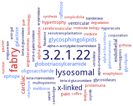
Word Map on EC 3.2.1.22
-
3.2.1.22
-
fabry
-
lysosomal
-
x-linked
-
glycosphingolipids
-
cardiac
-
globotriaosylceramide
-
endothelial
-
galactose
-
epitope
-
oligosaccharide
-
pain
-
raffinose
-
hypertrophy
-
ventricular
-
proteinuria
-
glycosidases
-
glycolipids
-
angiokeratomas
-
alpha-glucosidase
-
melibiose
-
cerebrovascular
-
galactomannans
-
simplicifolia
-
multisystemic
-
corporation
-
xenotransplantation
-
anaphylaxis
-
hemizygotes
-
cambridge
-
alpha-l-fucosidase
-
stachyose
-
coffee
-
hyperacute
-
anti-gal
-
beta-d-glucuronidase
-
analysis
-
bandeiraea
-
alpha-n-acetylgalactosaminidase
-
guar
-
beta-d-galactosidase
-
biotechnology
-
sanofi
-
nutrition
-
griffonia
-
beta-mannosidase
-
imiglucerase
-
degradation
-
gaucher
-
molecular biology
-
beta-d-glucosidase
-
medicine
-
synthesis
-
verticillata
-
soymilk
-
alpha-d-mannosidase
-
arylamidase
-
flatulence
- 3.2.1.22
- fabry
- lysosomal
-
x-linked
- glycosphingolipids
- cardiac
- globotriaosylceramide
- endothelial
- galactose
- epitope
- oligosaccharide
- pain
- raffinose
- hypertrophy
- ventricular
- proteinuria
- glycosidases
- glycolipids
- angiokeratomas
- alpha-glucosidase
- melibiose
- cerebrovascular
- galactomannans
- simplicifolia
-
multisystemic
-
corporation
-
xenotransplantation
- anaphylaxis
-
hemizygotes
-
cambridge
- alpha-l-fucosidase
- stachyose
- coffee
-
hyperacute
-
anti-gal
- beta-d-glucuronidase
- analysis
-
bandeiraea
- alpha-n-acetylgalactosaminidase
- guar
- beta-d-galactosidase
- biotechnology
-
sanofi
- nutrition
- griffonia
- beta-mannosidase
- imiglucerase
- degradation
- gaucher
- molecular biology
- beta-d-glucosidase
- medicine
- synthesis
-
verticillata
-
soymilk
- alpha-d-mannosidase
- arylamidase
-
flatulence
Reaction
Synonyms
1,6-alpha-D-galactoside galactohydrolase, a-galactosidase, Ag-I, Ag-II, Aga-F78, Aga-Y, AgaA, agaAJB13, AgaB, AgaI, AGal, AgalB, Agalsidase alfa, AglA, AglC, AkalphaGal, alkaline alpha-gal form 1, alkaline alpha-galactosidase, alkaline alpha-galactosidase form 1, alpha-D-galactopyranoside galactohydrolase, alpha-D-galactosidase, Alpha-D-galactoside galactohydrolase, alpha-Gal, alpha-Gal A, alpha-Gal II, alpha-Gal III, alpha-gal1, alpha-galactosidase, alpha-galactosidase 1, alpha-galactosidase 2, alpha-galactosidase 3, alpha-galactosidase A, alpha-galactosidase AgaA A355E, alpha-galactosidase AgaB, alpha-galactosidase I, alpha-galactosidase II, alpha-galactosidase III, alpha-galactoside galactohydrolase, alphaGal1, ATSIP2, blAga3, BLGA_00330, ceramidase, galactosylgalactosylglucosyl-, ceramide trihexosidase, ceramidetrihexosidase, ceramidetrihexoside-alpha-galactosidase, E1 alpha-galactosidase, E2 alpha-galactosidase, E3 alpha-galactosidase, EC 3.2.1.47, Fabrazyme, Gal36, GALA, galA17, GalS, Genzyme, GH36 alpha-galactosidase, GH97b, GLA, LaMel36A, MEL1, Mel4A, MelA, melibiase, retaining alpha-galactosidase, ScAGal, Tm GalA, TM1192, TmGalA, TnGalA, trihexosyl ceramide galactosidase, trihexosylceramide alpha-galactosidase, trihexosylceramidealpha-galactosidase
ECTree

Advanced search results
Crystallization
Crystallization on EC 3.2.1.22 - alpha-galactosidase
Please wait a moment until all data is loaded. This message will disappear when all data is loaded.
 top
topCRYSTALLIZATION (Commentary) 

ORGANISM
UNIPROT
LITERATURE
hanging drop vapor diffusion method, using 100 mM MES-NaOH buffer, pH 6.2, containing 12% (w/v) PEG (polyethylene glycol) 6000 or 100 mM MES-NaOH buffer containing 10% (w/v) PEG 10000, at 20°C
vapor diffusion method, using 80 mM sodium acetate, 20% (w/v) polyethylene glycol 4000, and 6.4% (v/v) isopropanol
vapour-diffusion method. Crystals of AgaA A355E belong to space group P3(1) 21 or P3(2) 21,with unit-cell parameters a = b = 150.1, c = 233.2 A
-
vapour-diffusion method. Crystals of AgaB belong to space group I222 or I2(1)2(1)2(1),with unit-cell parameters a = 87.5, b = 113.13, c = 161.6 A
-
construction of structural models of mutant enzyme responsible for Fabry disease and calculation of indexes. Structural changes in the classic Fabry disease group are generally large and tend to be in the core region of a protein or located in the functionally important region, including the active-site pocket. Structural changes in the variant Fabry disease group are small or localized on the surface of the molecule far away from the activte site. Structural changes due to amino acid substitutions for which substrate analogues are effective for improving the stability or transportation are small or localized on the molecular surface
-
hanging drop vapor diffusion method, using 25% (w/v) PEG 4000, 200 mM (NH4)2SO4, and 100 mM NaCH3COO, pH 4.6, at 20°C
the crystal structure of alpha-galactosidase from Lactobacillus acidophilus NCFM (LaMel36A) is determined by single-wavelength anomalous dispersion. In addition, a 1.58-A-resolution crystallographic complex with alpha-D-galactose at substrate binding subsite-1 is determined. LaMel36A has a largeN-terminal twisted beta-sandwich domain, connected by a long alpha-helix to the catalytic (beta/alpha)8-barrel domain, and a C-terminal beta-sheet domain
Q7WWP9
hanging-drop vapor diffusion method, crystal structure determined at 1.5 A resolution
-
crystal structures of tetrameric Saccharomyces cerevisiae alpha-galactosidase and its complexes with the substrates melibiose and raffinose have been determined to 1.95, 2.40, and 2.70 A resolution. The monomer folds into a catalytic (alpha/beta)8 barrel and a C-terminal beta-sandwich domain with unassigned function
in vitro deglycosylated protein, sitting drop vapor diffusion method, using 18-20% (w/v) PEG 3350, 0.1 M bis-tris propane pH 7.5-8.5, and 0.2 M KSCN
hanging-drop method, structure of alpha-galactosidase is determined at 1.54 A resolution and its complex with the competitive inhibitor beta-D-galactose refined at 2.0 A resolution
polyethylene glycol 4000 solution, hanging drop method, orthorhombic space group
-