4.2.3.146: cyclooctat-9-en-7-ol synthase
This is an abbreviated version!
For detailed information about cyclooctat-9-en-7-ol synthase, go to the full flat file.
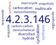
Word Map on EC 4.2.3.146
Reaction
Synonyms
cetB2, CotB2, CYC, diterpene cyclooctatin synthase
ECTree

Advanced search results
Crystallization
Crystallization on EC 4.2.3.146 - cyclooctat-9-en-7-ol synthase
Please wait a moment until all data is loaded. This message will disappear when all data is loaded.
 top
topCRYSTALLIZATION (Commentary) 

ORGANISM
UNIPROT
LITERATURE
purified recombinant native and SeMet-labeled enzyme CotB2 with bound substrate analogue geranylgeranyl thiodiphosphate, hanging drop vapor diffusion method, mixing of 0.001 ml of 10 mg/ml protein, in 20 mM Tris-HCl, pH 8.0, 5 mM MgSO4, with or without 1 mM substrate, with 0.001 ml of reservoir solution, and equilibration against 0.5 ml of reservoir solution, 20°C, the reservoir solution contains 0.1 M HEPES-NaOH, pH 7.0-8.5, and 1.4-1.5 M ammonium formate for the apoenzyme, 0.1 M Tris-HCl, pH 8.0, and 2.4 M ammonium formate for the apo-SeMet-enzyme, and 0.1 M bicine-NaOH, pH 9.0, and 20% PEG 6000 for the substrate-bound enzyme, X-ray diffraction structure determination and analysis. Molecular replacement. Only a single Mg2+ ion is present in the observed structure, coordinated by the Asn220, Ser224, and Glu228 side chains belonging to the canonical NSE/DTE motif