5.3.1.23: S-methyl-5-thioribose-1-phosphate isomerase
This is an abbreviated version!
For detailed information about S-methyl-5-thioribose-1-phosphate isomerase, go to the full flat file.
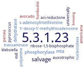
Word Map on EC 5.3.1.23
-
5.3.1.23
-
salvage
-
phosphorylase
-
mta
-
dehydratase
-
s-adenosylmethionine
-
ribose-1,5-bisphosphate
-
mill
-
5'-deoxy-5'-methylthioadenosine
-
aci-reductone
-
universally
-
avocado
-
persea
-
nucleosidase
-
auxotrophic
-
klebsiella
-
transaminase
-
pyrococcus
-
horikoshii
-
americana
-
polyamine
-
brewing
- 5.3.1.23
-
salvage
- phosphorylase
- mta
- dehydratase
- s-adenosylmethionine
-
ribose-1,5-bisphosphate
-
mill
- 5'-deoxy-5'-methylthioadenosine
-
aci-reductone
-
universally
- avocado
- persea
- nucleosidase
-
auxotrophic
-
klebsiella
- transaminase
-
pyrococcus
- horikoshii
- americana
- polyamine
- brewing
Reaction
Synonyms
1-PMTR isomerase, 5'-methylthioribose-1-phosphate isomerase, 5-methylthio-5-deoxy-D-ribose-1-phosphate ketol-isomerase, 5-methylthioribose 1-phosphate isomerase, 5-methylthioribose-1-phosphate isomerase, Bs-M1Pi, Isomerase, methylthioribose 1-phosphate, M1Pi, methylthioribose-1-phosphate isomerase, Meu1p, MRDI, MRI1, Mri1p, mtnA, MTR-1-P, MTR-1-P isomerase, PH0702, PhM1Pi, Ypr118w, Ypr118wp
ECTree

Advanced search results
Subunits
Subunits on EC 5.3.1.23 - S-methyl-5-thioribose-1-phosphate isomerase
Please wait a moment until all data is loaded. This message will disappear when all data is loaded.
 top
topSUBUNIT 

ORGANISM
UNIPROT
COMMENTARY 
LITERATURE
dimer
homodimer
additional information
additional information

enzyme structure and active site structure comparisons, open/closed conformational transition of enzyme M1Pi. The structure of Bs-M1Pi shows that the active site is completely shielded from the solvent region. The substrate binding is likely to induce the large conformational changes of N- and C-terminal domains as well as the rearrangement of the hydrogen-bond network around the loops 93-98 and 290-294 to stabilize the closed state of the enzyme
additional information
-
enzyme structure and active site structure comparisons, open/closed conformational transition of enzyme M1Pi. The structure of Bs-M1Pi shows that the active site is completely shielded from the solvent region. The substrate binding is likely to induce the large conformational changes of N- and C-terminal domains as well as the rearrangement of the hydrogen-bond network around the loops 93-98 and 290-294 to stabilize the closed state of the enzyme
-
additional information
enzyme three-dimensional structure analysis and structure comparisons. The most visible difference between the open and closed conformation can be perceived in the position of the loop connecting the helices alpha3 and alpha4 of the NTD. In the open state, the loop is away from the active-site pocket, whereas in the closed state the loop moves toward the active-site pocket with a forward shift of about 24 A