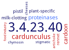
3.4.23.40: Phytepsin
This is an abbreviated version!
For detailed information about Phytepsin, go to the full flat file.
Word Map on EC 3.4.23.40
-
3.4.23.40
-
cardunculus
-
cardoon
-
proteinases
-
pistil
-
milk-clotting
-
plant-specific
-
rennet
-
stigmatic
-
artichoke
-
chymosin
-
cheeses
-
food industry
- 3.4.23.40
- cardunculus
- cardoon
- proteinases
- pistil
-
milk-clotting
-
plant-specific
- rennet
-
stigmatic
- artichoke
- chymosin
-
cheeses
- food industry
Reaction
Prefers hydrophobic residues Phe, Val, Ile, Leu, and Ala at P1 and P1', but also cleaves -Phe-/-Asp- and -Asp-/-Asp- bonds in 2S albumin from plant seeds =
Synonyms
Acid protease, Ap1, Aspartic proteinase, Aspartyl endoproteinase, Barley grain aspartic proteinase, Carboxyl proteinase, CardA, cardB, Cardosin, cardosin A, cardosin B, Cynarase, cynarase A, cynarase B, cynarase C, Edestinase, HvAP, Nepenthesin, Oryzasin, PCB, procardosin B
ECTree

Advanced search results
Crystallization
Crystallization on EC 3.4.23.40 - Phytepsin
Please wait a moment until all data is loaded. This message will disappear when all data is loaded.
 top
topCRYSTALLIZATION (Commentary) 

ORGANISM
UNIPROT
LITERATURE
to 1.72 A resoultion. Structure includes two molecules, built up by two glycosylated peptide chains of 31 and 15 kDa. The glycosyl content is described by 19 sugar rings attached to residues Asn67 and Asn257 localized on the molecular surface away from the conserved active site. A hydrogen bond between Gln126 and Manbeta4 renders the monosaccharide oxygen O2 sterically inaccessible to accept a xylosyl residue, explaining a hitherto unknown type of plant glycan. The Arg-Gly-Asp sequence, involved in recognition of a putative cardosin A receptor, is found in a loop between two beta-strands on the molecular surface opposite the active site cleft
analysis of solvent structure in crystals, comparison of aspartic proteases, zymogen form
-