5.4.3.8: glutamate-1-semialdehyde 2,1-aminomutase
This is an abbreviated version!
For detailed information about glutamate-1-semialdehyde 2,1-aminomutase, go to the full flat file.
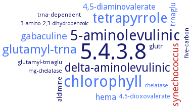
Word Map on EC 5.4.3.8
-
5.4.3.8
-
chlorophyll
-
5-aminolevulinic
-
tetrapyrrole
-
glutamyl-trna
-
delta-aminolevulinic
-
synechococcus
-
gabaculine
-
hema
-
4,5-diaminovalerate
-
trnaglu
-
4,5-dioxovalerate
-
aldimine
-
glutr
-
five-carbon
-
glutamyl-trnaglu
-
trna-dependent
-
3-amino-2,3-dihydrobenzoic
-
chelatase
-
mg-chelatase
- 5.4.3.8
- chlorophyll
-
5-aminolevulinic
- tetrapyrrole
- glutamyl-trna
-
delta-aminolevulinic
- synechococcus
- gabaculine
- hema
- 4,5-diaminovalerate
- trnaglu
- 4,5-dioxovalerate
-
aldimine
- glutr
-
five-carbon
-
glutamyl-trnaglu
-
trna-dependent
-
3-amino-2,3-dihydrobenzoic
- chelatase
-
mg-chelatase
Reaction
Synonyms
Aminotransferase, glutamate semialdehyde, AtGSA1, EC 2.7.2.13, Glutamate 1-semialdehyde aminotransferase, glutamate-1-semialdehyde amino-transferase, glutamate-1-semialdehyde aminomutase, Glutamate-1-semialdehyde aminotransferase, glutamate-1-semialdehyde-2,1-aminomutase, glutamate-1-semialdehyde-aminomutase, GSA, GSA aminotransferase, GSA-AT, GSA1, GSAM, GSAT, HemL, PA4088, protein PA4088
ECTree

Advanced search results
Subunits
Subunits on EC 5.4.3.8 - glutamate-1-semialdehyde 2,1-aminomutase
Please wait a moment until all data is loaded. This message will disappear when all data is loaded.
 top
topSUBUNIT 

ORGANISM
UNIPROT
COMMENTARY 
LITERATURE
dimer
additional information
dimer

enzyme AtGSA1 forms an asymmetric dimer and displays asymmetry in cofactor binding as well as in the gating-loop orientation
dimer
three-dimensional structure analysis of wild-type and mutant enzymes, overview
dimer
Pseudomonas aeruginosa ATCC 15692 / DSM 22644 / CIP 104116 / JCM 14847 / LMG 12228 / 1C / PRS 101 / PAO1
-
three-dimensional structure analysis of wild-type and mutant enzymes, overview
-
additional information
the overall structure of AtGSA1 consists of three sequentially arranged domains: the N-terminal domain (Val1-Asp63, mature protein) comprises one alpha-helix and a three-stranded antiparallel beta-sheet, the pyridoxamine 5'-phosphate/pyridoxal 5'-phosphate-binding domain (Tyr64-Gly328), which is also the catalytic domain, contains a central seven-stranded beta-sheet with one antiparallel and six parallel beta-strands, and the C-terminal domain (Thr329-Ile434) is composed of a three-stranded antiparallel beta-sheet with four helices covering the outer surface. Asymmetry of AtGSA1 in the gating-loop conformation, overview
additional information
-
the overall structure of AtGSA1 consists of three sequentially arranged domains: the N-terminal domain (Val1-Asp63, mature protein) comprises one alpha-helix and a three-stranded antiparallel beta-sheet, the pyridoxamine 5'-phosphate/pyridoxal 5'-phosphate-binding domain (Tyr64-Gly328), which is also the catalytic domain, contains a central seven-stranded beta-sheet with one antiparallel and six parallel beta-strands, and the C-terminal domain (Thr329-Ile434) is composed of a three-stranded antiparallel beta-sheet with four helices covering the outer surface. Asymmetry of AtGSA1 in the gating-loop conformation, overview