4.2.2.11: guluronate-specific alginate lyase
This is an abbreviated version!
For detailed information about guluronate-specific alginate lyase, go to the full flat file.
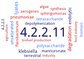
Word Map on EC 4.2.2.11
-
4.2.2.11
-
lyases
-
degradation
-
klebsiella
-
unsaturated
-
polysaccharide
-
endolytic
-
pneumoniae
-
depolymerization
-
synthesis
-
aerogenes
-
flavobacterium
-
4.2.2.3
-
tetrasaccharide
-
algae
-
terrestrial
-
mannuronate
-
sphingomonas
-
biomass
-
biofuel production
-
agriculture
-
industry
-
food industry
- 4.2.2.11
- lyases
- degradation
-
klebsiella
- unsaturated
- polysaccharide
-
endolytic
- pneumoniae
-
depolymerization
- synthesis
- aerogenes
- flavobacterium
-
4.2.2.3
- tetrasaccharide
- algae
-
terrestrial
-
mannuronate
- sphingomonas
- biomass
- biofuel production
- agriculture
- industry
- food industry
Reaction
Synonyms
A1m, A9mC, A9mL, A9mT, Alg17C, Alg2A, Alg7D, AlgB, alginase II, alginate (poly-alpha-L-guluronate) lyase, alginate lyase, AlgL, AlgL5, AlgMytC, alkaliphilic alginate lyase, ALY, Aly-SJ02, Aly1, Aly5, AlyA, AlyA1PL7, AlyA5, AlyD, AlyDW11, AlyE, AlyQ, AlyV5, AXE80_11190, endo-type alginate lyase, guluronate lyase, KJ-2 alginate lyase, L-guluronan lyase, L-guluronate lyase, lyase, polyguluronate, Oal17A, oligoalginate lyase, poly(1,4-alpha-L-guluronide)lyase, poly(alpha-L-guluronate) lyase, poly-alpha-L-guluronate lyase, polyguluronate-specific alginate lyase, polyMG-specific alginate lyase, V12B01_09446, V12B01_24274, WP_053404615, xadAly7B
ECTree

Advanced search results
Crystallization
Crystallization on EC 4.2.2.11 - guluronate-specific alginate lyase
Please wait a moment until all data is loaded. This message will disappear when all data is loaded.
 top
topCRYSTALLIZATION (Commentary) 

ORGANISM
UNIPROT
LITERATURE
crystal structures of native protein and its inactive mutant H531A in complex with alginate trisaccharide, at 2.10 and 2.99 A resolutions with final R-factors of 18.3 and 19.9%, respectively. The enzyme is comprised of an alpha/alpha-barrel plus anti-parallel beta-sheet as a basic scaffold. His311 and Tyr365 are the catalytic base and acid, respectively. A short alpha-helix in the central alpha/alpha-barrel domain and a conformational change at the interface between the central and C-terminal domains are essential for the exolytic mode of action
20°C, drop solution comprising 1.4 M NaCl, 0.1 M potassium sodium phosphate and 0.1 M 2-morpholinoethanesulfonate-sodium hydroxide pH 6.5, vapor-diffusion method, monoclinic cristals
-
homology modeling of structure. Residues Asn198, His199, Arg246, and Tyr253 are conserved for the catalytic active site
purified recombinant soluble His-tagged wild-type and selenomethionine-labeled enzyme, and two mutant enzymes H202L and Y258A, and Y258A DELTAMMG variant, free or in complex with an alginate trisaccharide, hanging drop vapour diffusion method, mixing of 15 mg/ml protein in in 20 mM HEPES, pH 7.5, 100 mM KCl, with 0.1 M Tris, pH 8.0, 5% 2-methyl-2,4-pentanediol, 10% PEG 6000, 3 days, 16°C, X-ray diffraction structure determination and analysis at 1.85 A, 1.7 A, 2.45 A, and 1.9 A resolution, respectively
hanging-drop vapour-diffusion, A1-II structure at 2.2 A resolution, A1-II' structure at 1.0 A resolution
-
purified recombinant catalytic domain of AlyA1PL7, hanging drop vapor diffusion method, 0.002 ml of 11.3 mg/ml protein solution with 0.1 mg/ml oligoguluronate, are mixed with 0.001 ml of reservoir solution containing 0.2 M KSCN and 28% PEG-MME 2000, and equilibration against 0.5 ml reservoir solution, 21°C, screening and method optimization, X-ray diffraction structure determination and analysis at 1.43 A resolution
purified recombinant full-length dimeric AlyA5, hanging drop vapor diffusion method, 0.002 ml of 7.7 mg/ml protein solution with 0.1 mg/ml oligoglucuronate, are mixed with 0.001 ml of reservoir solution containing PEG 3350 and 0.2 M sodium/potassium tartrate, and equilibration against 02 ml reservoir solution, 21°C, screening and method optimization, X-ray diffraction structure determination and analysis at 1.75 A resolution