4.2.1.1: carbonic anhydrase
This is an abbreviated version!
For detailed information about carbonic anhydrase, go to the full flat file.
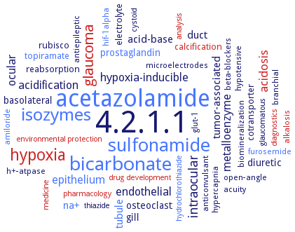
Word Map on EC 4.2.1.1
-
4.2.1.1
-
acetazolamide
-
bicarbonate
-
sulfonamide
-
isozymes
-
hypoxia
-
glaucoma
-
intraocular
-
acidosis
-
epithelium
-
hypoxia-inducible
-
metalloenzyme
-
endothelial
-
ocular
-
acidification
-
acid-base
-
na+
-
tubule
-
duct
-
tumor-associated
-
prostaglandin
-
diuretic
-
osteoclast
-
basolateral
-
gill
-
rubisco
-
calcification
-
topiramate
-
reabsorption
-
cotransporter
-
electrolyte
-
glut-1
-
branchial
-
open-angle
-
antiepileptic
-
beta-blockers
-
h+-atpase
-
acuity
-
hif-1alpha
-
hypotensive
-
anticonvulsant
-
hypercapnia
-
alkalosis
-
microelectrodes
-
amiloride
-
furosemide
-
biomineralization
-
drug development
-
pharmacology
-
environmental protection
-
diagnostics
-
cystoid
-
thiazide
-
medicine
-
hydrochlorothiazide
-
analysis
-
glaucomatous
- 4.2.1.1
- acetazolamide
- bicarbonate
- sulfonamide
- isozymes
- hypoxia
- glaucoma
-
intraocular
- acidosis
- epithelium
-
hypoxia-inducible
-
metalloenzyme
- endothelial
-
ocular
-
acidification
-
acid-base
- na+
- tubule
- duct
-
tumor-associated
- prostaglandin
-
diuretic
- osteoclast
-
basolateral
- gill
- rubisco
- calcification
- topiramate
-
reabsorption
-
cotransporter
-
electrolyte
-
glut-1
-
branchial
-
open-angle
-
antiepileptic
-
beta-blockers
- h+-atpase
-
acuity
- hif-1alpha
-
hypotensive
-
anticonvulsant
-
hypercapnia
- alkalosis
-
microelectrodes
- amiloride
- furosemide
-
biomineralization
- drug development
- pharmacology
- environmental protection
- diagnostics
-
cystoid
-
thiazide
- medicine
- hydrochlorothiazide
- analysis
-
glaucomatous
Reaction
Synonyms
(CA) XIV, AaCA1, alkalistable alpha-carbonic anhydrase, alpha carbonic anhydrase, alpha class carbonic anhydrase, alpha-CA, alpha-carbonic anhydrase, alpha-carbonic anhydrase isozyme II, alpha-class carbonic anhydrase, alpha-type CA, alpha-type carbonic anhydrase, alphaCA, alphaCA1, Am CA, AmCA, anhydrase, ASCA, AtCA1, AtCA2, AtCAL1, AtCAL2, B13-CA, BCA II, bCA IV, BCAII, BCAIIGln253Cys, beta carbonic anhydrase, beta-CA, beta-carbonic anhydrase, beta-class carbonic anhydrase, betaCA, betaCA1, BhCA, bovine carbonic anhydrase II, bsCA I, bsCA II, CA, CA 1, CA 3, CA I, CA II, CA III, CA IV, CA IX, CA VA, CA VB, CA VI, CA VII, CA VIII, CA XII, CA XIII, CA XIV, Ca XV, CA-I, CA-II, CA-III, CA-IX, CA-VA, CA-VB, CA-VI, CA-VII, CA-XII, CA-XIV, CA1, CA14x, CA2, CA2 homolog, CA2-like, CA3, CA4, CA5A, CA6, CA9, CAA, CAA1, CAA2, Cab, cab-type beta-class carbonic anhydrase, cadmium carbonic anhydrase, CAH3, CAH7, CAH8, CAI, CAII, CAIII, CAIV, CAIX, Cam, CamH, CaN, Can2, canA, canB, CaNce103, carbonate anhydrase, Carbonate dehydratase, carbonate dehydratase I, carbonate dehydratase III, Carbonate dehydratase IX, Carbonate dehydratase VA, Carbonate dehydratase VB, Carbonate dehydratase VI, Carbonate dehydratase VII, Carbonate dehydratase XII, Carbonate dehydratase XIV, carbonate hydro-lyase, carbonate hydrolase, carbonate hydrolyase, carbonic acid anhydrase, carbonic anhydrase, carbonic anhydrase 1, carbonic anhydrase 14, carbonic anhydrase 2, carbonic anhydrase 3, carbonic anhydrase 5A, carbonic anhydrase 6, carbonic anhydrase 9, carbonic anhydrase cambialistic enzyme, carbonic anhydrase I, carbonic anhydrase I (CA I) Michigan 1, carbonic anhydrase II, carbonic anhydrase III, carbonic anhydrase isozyme I, carbonic anhydrase isozyme II, carbonic anhydrase isozyme III, carbonic anhydrase isozyme IV, carbonic anhydrase isozyme IX, carbonic anhydrase IV, carbonic anhydrase IX, carbonic anhydrase type III, carbonic anhydrase V, carbonic anhydrase VA, carbonic anhydrase VI, carbonic anhydrase VII, carbonic anhydrase XII, carbonic anhydrase XIII, carbonic anhydrase XIV, carbonic anhydrase XV, carbonic anhydrase-I, carbonic anhydrase-II, carbonic anhydrase-related protein, carbonic anhydrase-related protein VIII, carbonic dehydratase, carboxyanhydrase, CARP, CAS1, CAS2, CasCAc, CasCAg, CAV, CAVA, CAXII, CcaA, CcmM, CDCA1, chloroplast carbonic anhydrase, clCA, CmCA, CO2 hydrase, CO2 hydratase, CPB, CpsCA, CpsCAgamma, CrCAH3, CsoSCA, cynT, CynT2, cytoplasmic carbonic anhydrase, Dca, dCA I, dCA II, dCAII, DDCA, dehydratase, carbonate, delta carbonic anhydrase, delta-carbonic anhydrase, delta-class CA, diatom carbonic anhydrase, Dsp-aCAopt sp., dual domain-carbonic anhydrase, dual-domain carbonic anhydrase, DVU_1777, E84A MTCA, eCA, ECCA, erythrocyte carbonic anhydrase, eta-CA, eta-carbonic anhydrase, external carbonic anhydrase, extracellular carbonic anhydrase, FbiCA 1, fCA, gamma carbonic anhydrase 1, gamma carbonic anhydrase 2, gamma carbonic anhydrase-like 1, gamma carbonic anhydrase-like 2, gamma-CA, gamma-CA1, gamma-CA2, gamma-CAL1, gamma-CAL2, gamma-carbonic anhydrase, gamma-class carbonic anhydrase, gammaCA, H216N ATCA, H64A HCA II, HC II, HCA, hCA I, HCA II, hCA III, hCA IV, hCA IX, hCA VA, hCA VB, hCA VI, hCA VII, hCA XII, hCA XIV, hCA XIV catalytic domain, HCA-I, HCA-II, hCAII, HcCA3, HICA, HP1186, hpalphaCA, hpbetaCA, human carbonic anhydrase, human carbonic anhydrase I, human carbonic anhydrase II, human carbonic anhydrase III, human carbonic anhydrase isoenzyme I, human carbonic anhydrase isoenzyme II, human carbonic anhydrase IX, human carbonic anhydrase XII, human carbonic anhydrase XIV, Ice-CA, icfA, isozyme CA II, isozyme CA IV, LdcCA, luminal carbonic anhydrase, lwCA, mCA V, mCA XIII, Membrane antigen MN, mesohalophilic carbonic anhydrase, MG-CA, MgaCA, More, mtcA1, mtcA2, MtCam, MTCY373.03, Nce103, NcoCA, NGCA, NstCcmM209, OEC33 protein, P54/58N, pentraxin-CA VI, pentraxin-carbonic anhydrase, PERMA_1443, PF3D7_1140000, PfCAdom, PgiCA, PgiCAb, PGJ_00014320, PhaCAgamma, PhCamH, photosystem II-associated carbonic anhydrase, Pl-can, plant-type (beta-class) carbonic anhydrase, PMCA, pMW1, poly-[Hb-SOD-CAT-CA], polyhemoglobin-superoxide dismutase-catalase-carbonic anhydrase, PtCA1, RCC-associated antigen G250, Renal cell carcinoma-associated antigen G250, RT erythrocyte CA, Rv1248, Rv1284, Rv3273, Rv3588c, SABP3, Salivary carbonic anhydrase, SazCA, scCA, Secreted carbonic anhydrase, secretory carbonic anhydrase, secretory carbonic anhydrase VI, SspCA, STPCA, STPCA-2, SULAZ_0541, TacA, TCAb, TCAc, TcCA, Tcru, Tcr_1545, Theam_1576, tobacco salicylic acid-binding protein 3, Tumor antigen HOM-RCC-3.1.3, TWCA1, TweCA, VchCA
ECTree

Advanced search results
Crystallization
Crystallization on EC 4.2.1.1 - carbonic anhydrase
Please wait a moment until all data is loaded. This message will disappear when all data is loaded.
 top
topCRYSTALLIZATION (Commentary) 

ORGANISM
UNIPROT
LITERATURE
crystals belong to hexagonal space group P6(1), with unit-cell parameters a : b : 66.7 A, c : 240.0 A
-
X-ray absorption near edge spectroscopy (XANES)/MXAN analysis of the reaction center
purified native isozyme alphaCA1, 0.002 ml of 20 mg/ml protein in 25 mM Tris-HCl, pH 7.5, is mixed with 0.002 ml of reservoir solution, containing 1.0 M ammonium sulfate and 100 mM Tris-HCl, pH 8.5, and equilibrated against 0.7 ml of reservoir solution, 20°C, X-ray diffraction structure determination and analysis at 1.88 A resolution, MAD method
-
purified recombinant detagged enzyme CrCAH3 with inhibitors phosphate and acetazolamide, hanging drop vapor diffusion method, mixing of 0.001 ml of 3.6 mg/l protein in mM Tris-HCl, pH 8.0, and 150 mM NaCl, and 1 mM ligand, with 0.001 ml of reservoir solution containing 2.5 M NH4H2PO4 and 0.1 M Tris-HCl, pH 8.0, to a final pH of pH 4.1, and equilibration over 1 ml reservoir solution, at 18°C, method optimization, X-ray diffraction structure determination and analysis at 2.6-2.7 A resolution
crystal structure of dCA II at 1.86 A resolution, vapour diffusion method
-
crystallized by the hanging drop vapour-diffusion method, monoclinic crystal system, crystals belong to space group P2(1), with unit-cell parameters a : 47.0 A, b : 119.9 A, c : 58.5 A
-
2 different tetragonal crystal forms, form 1 space group P4(2)2(1)2, unit cell dimensions a : b : 68.54 A, c : 85.88A, form 2 space group P4(3)22, a : : b : 81.24 A, c : 162.14 A
CynT2, form 1, space group P4(2)212, unit-cell parameters a : b : 68.54 A, c : 85.88 A, form 2, space group P4(3)22, a : b : 81.00 A, c : 161.98 A, form 3, space group P2(1), a : 48.21, b : 140.73, c : 72.57, selenomethionine-substituted crystals, space group P4(3)22, a : b : 81.24 A, c : 162.14 A
purified recombinant Co-HICA by hanging drop vapor diffusion, 10 mg/ml protein crystallized in 0.2 M sodium acetate, 0.1 M Tris-HCl, pH 8.5, 0.1 M (NH4)2SO4, and 27% PEG 4000, 22°C, several days, X-ray diffraction structure determination and analysis at 2.5 A resolution
-
purified wild-type and mutant enzymes, 12 mg/ml protein mixed with 0.7 M sodium potassium tartrate, 0.10 M HEPES, pH 7.5, for tetragonal crystals and with 1.8 M ammonium sulfate, 4% PEG 400, 0.10 M HEPES, pH 7.5, for monoclinic crystals, 22°C, 2-3 days, crystals are soaked for 1-2 min in artificial mother liquor plus either 30% glucose or 30% glucose and 100 mM NaHCO3, X-ray diffraction structure determination and analysis
hanging drop vapor diffusion method. 2.2 A crystal structure of CsoSCA
-
all variants crystallized in the orthorhombic P2(1) 2(1) 2 (1) space group and are highly isomorphous with approximate unit cell dimensions a=42, b=72 and c=75 A and diffract between 1.56 and 2.0 A resolution
CA XII crystallized by the hanging drop vapor diffusion method, space group C2 with unit cell dimensions a : 146.7 A, b : 44.6 A, c : 85.2 A
carbonic anhydrase II complexed with two aromatic sulfonamide inhibitors N-(4-sulfamoylphenyl)-2-(thiophen-2-yl)acetamide and N-(2-fluoro-4-sulfamoylphenyl)-2-(thiophen-2-yl)acetamide incorporating 2-thienylacetamido moieties, hanging drop vapor diffusion method, drops of 0.005 ml containing 0.5 mM hCA II, 1 mM inhibitor, 0.1% DMSO, 0.8 M sodium citrate, 50 mM Tris-HCl, pH 8.0,are equilibrated against 1 ml precipitant solution containing 1.6 M sodium citrate and 50 mM Tris-HCl, pH 8.0, at room temperature, X-ray diffraction structure determination and analysis at 1.6-1.7 A resolution
-
crystal structure of the hCA II/2-N,N-dimethylamino-1,3,4-thiadiazole-5-methanesulfonamide adduct
crystal structure of the hCA/2-(hydrazinocarbonyl)-3-phenyl-1H-indole-5-sulfonamide adduct
crystal structures of C-terminal hexahistidine-tagged carbonic anhydrase III and F198L carbonic anhydrase III, hanging-drop vapour diffusion method, 2.1 A resolution
crystals obtained by hanging drop technique, hCA II-sulfamate, space group P2(1), a : 42.32 A, b : 41.48 A, c : 72.44 A, hCA II-sulfamide, space group P2(1), a : 42.70 A, b : 41.70 A, c : 73.00 A
crystals obtained by hanging drop technique, space group P2(1), cell parameters a: 42.01 A, b : 41.43 A, c: 71.99 A
crystals obtained using the hanging drop vapor diffusion method, isozyme CA I Michigan I, space group P2(1)2(1)2(1), with unit cell dimensions parameters a : 62.50 A, b : 72.13 A, c : 121.54 A, isozyme CA I Michigan 1-Zn(II)2, space group P2(1)2(1)2(1), with unit cell dimensions parameters a : 62.68 A, b : 71.36 A, c : 120.91A
crystals of carbonic anhydrase II and carbonic anhydrase IX mimic are grown at room temperature using the hanging drop vapour diffusion method
crystals of K64H, R67H, and K64H/R67N HCA III are grown by hanging drop vapor diffusion method. X-ray crystal structures of site-specific mutants of human carbonic anhydrase III (HCA III): K64H, R67H, and K64H/R67N HCA III
crystals of the HCA II single-site mutants Y7F, N62L, and N67L are obtained using the hanging-drop vapor diffusion method. All crystals are isomorphous and belong to space group P2(1) with mean unit cell dimensions: a = 42.7 A, b = 41.6 A, c = 72.9 A and beta = 104.6°
hanging drop vapor diffusion method, using 0.4 M NH4H2PO4, 0.1 M sodium citrate, pH 5.0
holoenzyme and apoenzyme are crystallized by hanging drop vapour diffusion method, using 1.3 M sodium citrate, 100 mM Tris-HCl, pH 7.8
in complex with inhibitor RWJ-37497, crystals obtained by the hanging-drop vapour-diffusion method, space group P2(1) with unit cell parameters a : 42.28 A, b : 41.39 A, c : 72.51 A
isoform CA II in complex with 6,7-dimethoxy-3,4-dihydroisoquinoline-2(1H)-sulfonamide, hanging drop vapor diffusion method, using 2.5 M (NH4)2SO4, 0.3 M NaCl, 100 mM Tris-Cl, pH 8.2
isoform CA II, sitting drop vapor diffusion method, using 0.1 M sodium bicine, 0.2 M ammonium sulfate and 2 M sodium malonate
isozyme CA XIII in the unbound state and in complex with acetazolamide, hanging drop vapour diffusion method, in 20 mM Tris-HCl, pH 7.5, 150 mM NaCl, 1 mM dithiothreitol with 30% (w/v) polyethylene glycol 4000, 0.2 M ammonium acetate, 0.1 M sodium acetate, pH 4.6
-
mutant H64A, crystals produced by the hanging drop method, space group P2(1) with unit cell parameters of a : 42.5 A, b : 41.6 A, c : 72.7 A
mutants are crystallized by hanging drop vapour diffusion method, with 50 mM Tris-HCl (pH 9.0) and 1.3 M sodium citrate at 20°C
purified enzyme in complex with inhibitors (S)-4-(4-acetyl-3-benzylpiperazine-1-carbonyl)benzene-1-sulfonamide and (S)-4-[2-benzyl-4-(4-sulfamoylbenzoyl)piperazine-1-carbonyl]benzene-1-sulfonamide, sitting drop vapor diffusion method, mixing of 0.002 ml of 10 mg/ml protein in 20 mM Tris-HCl, pH 9.0, with 0.002 ml of a solution of 28-31% PEG 4000, 0.2 M sodium acetate, 0.1 M Tris, pH 8.5-9.0 and equilibration against the same solution at 23°C, fifteen days, afterwards the crystals are soaked in 5 mM inhibitor solutions for 3 days, X-ray diffraction structure determination and analysis at 1.5-1.6 A resolution
purified isozyme CA II (chimeric CA IX mimic) mutant in complex with inhibitors 4-phenylbenzene-1-sulfonamide, 4-(2-methylphenyl)benzene-1-sulfonamide, 4-(3-formylphenyl)benzene-1-sulfonamide, and 4-(3-quinolinyl)benzene-1-sulfonamide, sitting drop vapor diffusion method, mixing of 300-500 nl of 20 mg/mL of CA II and 41 mg/ml of the CA IX mimic solution with crystallization solution containing 1.6 M sodium citrate, 50 mM Tris-HCl, pH 7.8, in a ratio of 3:5 or 1:1, 25°C, 1 week, X-ray diffraction structure determination and analysis at 1.55-1.69 A resolution (PDB IDS 5SZ0, 5SZ1, 5SZ2, and 5SZ3), molecular replacement using the enzyme structure PBD ID 3KS3 without zinc and solvent as search model
purified isozyme CA IX in complex with inhibitors 4-phenylbenzene-1-sulfonamide, 4-(2-methylphenyl)benzene-1-sulfonamide, 4-(3-formylphenyl)benzene-1-sulfonamide, and 4-(3-quinolinyl)benzene-1-sulfonamide, X-ray diffraction structure determination and analysis at 1.15-1.78 A resolution (PDB IDS 5SZ4, 5SZ5, 5SZ6, and 5SZ7), molecular replacement using the enzyme structure PBD ID 3KS3 without zinc and solvent as search model
purified recombinant differently isotope-labeled isozyme hCAII, 14 mg/ml protein, both vapour diffusion and microbatch under-oil techniques are valuated, hanging and sitting drop set-ups with 0.01 ml drops, microseeding, deuteration of hCA II does not affect the crystal structure. Large, single crystals grow for both H and D enzyme versions in microbatch under-oil experiments at pH 8.5, room temperature, X-ray diffraction structure determination and analysis
purified recombinant differently isotope-labeled isozyme hCAIX, 17 mg/ml protein, both vapour diffusion and microbatch under-oil techniques are valuated, hanging and sitting drop set-ups with 0.01 ml drops, microseeding, room temperature, deuteration of hCA XI does not affect the crystal structure, but for D-hCA IX mimic no crystals appear in any of the under-oil batch trays at all pHs tested, while H-hCA IX SV gives several good crystals per drop at pH 8.5. X-ray diffraction structure determination and analysis
purified recombinant HCA II, crystals are prepared at pH 6.0, pH 8.5, and pH 11.0, hanging drop vapor diffusion method, crystal preparation from apo-Co(II)-HCA II by mixing 0.005 ml of protein solution with 0.005 ml of precipitant solution, containing 1.4 M sodium citrate, and 100 mM Tris-HCl, pH 9.0, at room temperature, and soaking of the crystals in 100 mM CoCl2, 1.4 M sodium citrate, 50 mM Tris-Cl, 50 mM 3-(cyclohexylamino)propanesulfonic acid with pH values of 6.0, 8.5 and 11.0, respectively, 2-3days, X-ray diffraction structure determination and analysis at 1.5-1.6 A resolution
purified recombinant human CA II in complex with imidazole-based activator CAA12i, hanging drop vapor diffusion method, mixing of 10 mg/ml protein and 0.2 mM ligand in 50 mM Tris-HCl, pH 7.8, with reservoir solution, containing 50 mM Tris HCl and 1.3 M sodium citrate, pH 7.4, in a 1:1 ratio, 23°C, 7 days without agitation, X-ray diffraction structure determination and analysis at 1.6 A resolution
purified recombinant K170 HCA II variants, hanging drop vapour diffusion method, 2-3days, X-ray diffraction structure determination at room temperature and analysis
-
purified recombinant wild-type enzyme and mutants M1-M4, X-ray diffraction structure determination and analysis at resolutions of 1.62-2.29 A
site-specific mutant Y7H, crystals produced by the hanging drop method, space group P2(1) with unit cell dimensions a : 42.9 A, b: 41.6 A, c : 73.0 A
sitting drop vapor diffusion method, using 10% (w/v) PEG 6000, 0.3 M NH4Cl pH 6.3, and 10% (v/v) ethylene glycol
the hCA II-sulthiame complex is crystallized by cocrystallization procedures. High resolution crystal structure of the hCA II-sulthiame adduct
the hCA IIN-hydroxysulfamide complex is cocrystallized at 4°C by the hanging drop vapor diffusion method. X-ray crystal structure of the human hCA II-N-hydroxysulfamide adduct
X-ray absorption nearedge spectroscopy (XANES)/MXAN analysis of the reaction center
X-ray crystallographic structure of the disulfide-containing HCAII (dsHCAII) is solved to 1.77 A resolution
purified recombinant enzyme, sitting drop vapour diffusion, using a precipitant solution consisting of 2% v/v Tacsimate, pH 4.0, 0.1 M sodium acetate trihydrate, pH 4.6, and 16% w/v PEG 3350, 10 days at 17°C, X-ray diffraction structure determination and analysis at 2.6 A resolution
cubic crystals, space group P2(1)3, Zn-Cam, unit cell a : b : c: 82.64 A, Zn-Cam-HCO3-, unit cell a : b : c: 82.51 A, Zn-Cam-SO42-, unit cell a : b : c: 82.68 A, Co-Cam, unit cell a : b : c: 82.34 A, Co-Cam-HCO3-, unit cell a : b : c: 82.58 A, Co-Cam-SO42-, unit cell a : b : c: 82.61 A
purified enzyme in complex with Zn2+ and Co2+, X-ray diffraction structure determination and analysis, PDB ID 1QRG
-
the crystal structure of the enzyme is determined at 2.8 A resolution using the method of multiple isomorphous replacement in combination with 3fold non-crystallographic symmetry averaging and phase extension
crystals grown by hanging drop method, orthorhombic space group P2(1)2(1)2(1) with unit cell dimensions a : 54.9 A, b : 113.2 A, c : 156.2 A
-
enzyme crystal structure determination and analysis, Cab dimer, PDB ID 1G5C
crystallized by hanging drop vapor diffusion method, crystals belong to space group P2(1) with unit cell dimesions a : 59.0 A, b : 75.6 A, c : 73.2 A
-
crystallized by the vapor diffusion method, hanging drop, space group C2, unit cell parameters a : 99.3 A, b : 66.7 A, c : 92.6 A
-
structure of the extracellular domain of murine CA XIV solved at 2.8 A resolution, and the enzyme-acetazolamide complex at 2.9 A resolution
in complex with the inhibitor acetic acid, crystals of the orthorhombic space group C222 with cell parameters a : 136.9 A, b : 143.3 A, c : 202.1 A
-
in complex with Ca2+, sitting drop vapour diffusion method, with 22.5% PEG 4000 in MES-NaOH pH 5.8 and 0.1 M lithium chloride as the precipitating agent
Nce103 in complex with a substrate analog at 2.04 A resolution, hanging drop vapor diffusion method, using 20% (w/v) PEG4000, 0.1 M sodium citrate, pH 5.6, 0.1 M sodium acetate, 20% (v/v) ethylene glycol, at 16°C
purified recombinant His-tagged enzyme incomplex with inhibitor acetazolamide, hanging drop vapor diffusion method, mixing of 0.001 ml of 8 mg/ml protein in 20 mM Tris, pH 8.3, and a 10-molar excess of the inhibitor with 0.001 m of reservoir solution containing 30% w/v PEG monomethyl ether 550, 0.05 M MgCl2, and 0.1 M HEPES, pH 7.5, and equilibration against 0.5 ml of reservoir solution, 1 week, room temperature, X-ray diffraction structure determination and analysis at 1.95 A resolution, molecular replacement using the structure of enzyme SspCA from Sulfurihydrogenibium yellowstonensis (PDB ID 4G7A) as a search model
purified enzyme in complex with inhibitor acetazolamide, X-ray diffraction structure determination and analysis, PDB ID 4G7A
purified recombinant truncated enzyme CcaA220, by sitting drop vapor diffusion method, mixing of 0.001 ml of 7 mg/ml protein solution with 0.001 ml of well solution containing 2.0 M Na formate and 0.1 M Na acetate, pH 4.6, X-ray diffraction structure determination and analysis at 1.45 A resolution, molecular replacement using the structure of a catalytic dimer (residues 121-318) from Pisum sativum beta-CA (PDB ID 1ekj) as a search model
Synechocystis sp. PCC 6803 / Kazusa
vapor diffusion method, using 1.0 M ammonium sulfate, 0.1 M [bis(2-hydroxyethyl)amino]tris(hydroxymethyl)methane, 1% (w/v) PEG 3350, pH 5.5
Thermosynechococcus vestitus
-