3.5.1.98: histone deacetylase
This is an abbreviated version!
For detailed information about histone deacetylase, go to the full flat file.
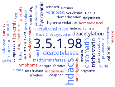
Word Map on EC 3.5.1.98
-
3.5.1.98
-
hdacs
-
chromatin
-
deacetylases
-
trichostatin
-
deacetylation
-
hydroxamic
-
valproic
-
saha
-
acetyltransferase
-
leukemia
-
butyrate
-
hyperacetylation
-
methyltransferase
-
vorinostat
-
repressor
-
lymphoma
-
microtubule
-
cyclin
-
sirtuins
-
nucleosome
-
t-cells
-
transactivation
-
caspase
-
bcl-2
-
metastasis
-
myeloid
-
proteasome
-
hypermethylation
-
tumorigenesis
-
cardiac
-
sirt1
-
neurodegenerative
-
hematological
-
prostate
-
demethylation
-
antiproliferative
-
corepressors
-
sirnas
-
cpg
-
non-histone
-
5-aza-2'-deoxycytidine
-
valproate
-
medicine
-
polycomb
-
bortezomib
-
dnmt3a
-
heterochromatin
-
creb-binding
-
epigenome
-
drug development
-
coactivator
-
relapsed
- 3.5.1.98
- hdacs
- chromatin
- deacetylases
-
trichostatin
-
deacetylation
-
hydroxamic
-
valproic
- saha
- acetyltransferase
- leukemia
- butyrate
-
hyperacetylation
- methyltransferase
- vorinostat
- repressor
- lymphoma
- microtubule
- cyclin
- sirtuins
- nucleosome
- t-cells
-
transactivation
-
caspase
- bcl-2
- metastasis
- myeloid
- proteasome
-
hypermethylation
- tumorigenesis
- cardiac
- sirt1
- neurodegenerative
- hematological
- prostate
-
demethylation
-
antiproliferative
-
corepressors
- sirnas
- cpg
-
non-histone
- 5-aza-2'-deoxycytidine
- valproate
- medicine
-
polycomb
- bortezomib
- dnmt3a
- heterochromatin
-
creb-binding
-
epigenome
- drug development
-
coactivator
-
relapsed
Reaction
hydrolysis of an N6-acetyl-lysine residue of a histone to yield a deacetylated histone =
Synonyms
acetyl-lysine deacetylase, AhHDA1, AtHD1, class I acetyl-lysine deacetylase, class I HDAC, class I histone deacetylase, class II Hda1 HDAC, class II histone deacetylase, class IIa HDAC, class IIa histone deacetylase, Clr6, cytoplasmic deacetylase, deacetylase-like amidohydrolase, dRPD3, FB188 HDAH, Glyma.01 g245100, Glyma.04 g000200, Glyma.04 g187000, Glyma.04 g187100, Glyma.05 g012900, Glyma.05 g021400, Glyma.05 g040600, Glyma.05 g192600, Glyma.06 g000100, Glyma.06 g178700, Glyma.11 g000300, Glyma.11 g187800, Glyma.12 g086700, Glyma.12 g188200, Glyma.17 g078000, Glyma.17 g085700, Glyma.17 g120900, Glyma.17 g229600, GmHDA1, GmHDA10, GmHDA11, GmHDA12, GmHDA13, GmHDA14, GmHDA15, GmHDA16, GmHDA17, GmHDA18, GmHDA2, GmHDA3, GmHDA4, GmHDA5, GmHDA6, GmHDA7, GmHDA8, GmHDA9, GmHDAC, HD1B, HD2, HD8, HDA, HDA-1, HDA-2, HDA-3, HDA-4, hda1, HDA15, HDA19, HDA9, HdaA, HDAC, HDAC 11, HDAC 3, HDAC1, HDAC10, HDAC2, HDAC2-1, HDAC2-2, HDAC3, HDAC4, HDAC5, HDAC6, HDAC6p114, HDAC6p131, HDAC7, HDAC8, HDAC9, HDAH, HDLP, HDT1, HDT2, histone deacetylase, histone deacetylase 1, histone deacetylase 10, histone deacetylase 11, histone deacetylase 2, histone deacetylase 3, histone deacetylase 4, histone deacetylase 6, histone deacetylase 7, histone deacetylase 8, histone deacetylase-1, histone deacetylase-7, histone deacetylase-like amidohydrolase, histone deacetylase-like protein, histone deacetylase1, Hos2, HosB, PA3774, pfHDAC-1, RPD3, Set3 complex, Set3C, tubulin deacetylase, zinc-dependent histone deacetylase, Zn(II)-dependent deacetylase, Zn-HDAC
ECTree

Advanced search results
Crystallization
Crystallization on EC 3.5.1.98 - histone deacetylase
Please wait a moment until all data is loaded. This message will disappear when all data is loaded.
 top
topCRYSTALLIZATION (Commentary) 

ORGANISM
UNIPROT
LITERATURE
free enzyme and in complexes with inhibitors trichostatin A and suberoylanilide hydroxamic acid. Active site consists of a tubular pocket, a zinc-binding site and two D-H charge-relay systems
-
modeling studies using inhibitor 6-[(9H-fluoren-3-ylmethyl)(3-phenoxybenzyl)amino]-N-hydroxyhexanamide and histone deacetylase-like protein, PDB ID: 1C3R. Inhibitor fills the equivalent space at the protein surface and occupies the catalytic binding site, coordinating the fundamental zinc ion
complex of isoform HDAC9 with myocyte enhancer factor-2 and DNA. HDAC9 binds to a hydrophobic groove of the myocyte enhancer factor-2 dimer. General mechainsm, by which class II histone deacetylases are recruited by myocyte enhancer factor-2
-
docking studies of the representative compound 4 to the homology model of HDAC1 and the X-ray structures of HDAH, PDB 1ZZ1, and HDAC8, PDB 1T67
-
molecular modeling of isoform HDAC8 in complex with inhibitor N-hydroxy-4-(methyl[(5-pyridin-2-yl-2-thienyl)sulfonyl]amino)benzamide
molecular modelling of structure of isoform HDAC8 in complex with suberoylanilide hydroxamic acid and with 7-mercapto-N-phenylheptanamide
N-terminal glutamine-rich fragment, residues 62-153. The glutamine-rich domain folds into an alpha-helix that assembles as a tetramer lacking regularly arranged apolar residues and an extended hydrophobic core. Instead, the protein interfaces consist of multiple hydrophobic patches interspersed with polar interaction networks, wherein clusters of glutamines engage in extensive intra- and interhelical interactions. In solution, the tetramer undergoes rapid equilibrium with monomer and intermediate species
seven structures of wild-type HDAC8 and mutants in complex with inhibitors or substrate are determined to a resolution of 1.8 to 3.3 A
the crystal structures of the catalytic domain of HDAC7 and its complexes with suberoylanilide hydroxamic acid and trichostatin A are solved
crystal structure of PA3774 from Pseudomonas aeruginosa as ligand-free enzyme and as protein-ligand complexes with a trifluoromethylketone inhibitor and the reaction product acetate, and of inhibitor-free PA3774H143A mutant, the inhibitor-free PA3774Y313F mutant, and the PA3774Y313F mutant in complex with the highly selective hydroxamate inhibitor N-[1,1,2,2,3,3,4,4,5,5,6,6-dodecafluoro-7-(hydroxyamino)-7-oxoheptyl]benzamide, from 0.5 M K2HPO4, 0.5 M Na2HPO4, and 0.1 M (NH4)2SO4, pH 7.5, X-ray diffraction structure determination and analysis
Hda1 ARB2 domain, native and selenomethionine-labebeed protein, hanging drop vapour diffusion method, from 0.1 M sodium acetate trihydrate, pH 4.6, and 2.0 M sodium formate, 12°C, 1 day, X-ray diffraction structure determination and analysis at 2.7 A resolution, molecular replacement method and modelling