3.5.1.52: peptide-N4-(N-acetyl-beta-glucosaminyl)asparagine amidase
This is an abbreviated version!
For detailed information about peptide-N4-(N-acetyl-beta-glucosaminyl)asparagine amidase, go to the full flat file.
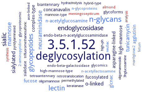
Word Map on EC 3.5.1.52
-
3.5.1.52
-
deglycosylation
-
n-glycans
-
lectin
-
glycopeptides
-
sialic
-
endoglycosidase
-
mannose
-
neuraminidase
-
o-linked
-
tunicamycin
-
sialylated
-
n-acetylglucosamine
-
asparagine-linked
-
fucose
-
glcnac
-
concanavalin
-
high-mannose
-
agglutinin
-
complex-type
-
fucosylated
-
sialidase
-
glycoforms
-
endo-beta-n-acetylglucosaminidase
-
biantennary
-
glycomics
-
asn-linked
-
high-mannose-type
-
glycoproteomic
-
n-acetyllactosamine
-
almond
-
tetraantennary
-
2-aminopyridine
-
meningosepticum
-
n-glycoproteins
-
endo-beta-galactosidase
-
fetuin
-
analysis
-
keratanase
-
permethylated
-
3hglucosamine
-
hybrid-type
-
mannose-type
-
oligomannosidic
-
synthesis
-
hydrazinolysis
-
man9glcnac2
-
trifluoromethanesulfonic
-
pyridylaminated
-
high-ph
-
oligomannose
-
retrotranslocation
-
neu5ac
- 3.5.1.52
-
deglycosylation
- n-glycans
- lectin
- glycopeptides
-
sialic
-
endoglycosidase
- mannose
- neuraminidase
-
o-linked
- tunicamycin
-
sialylated
- n-acetylglucosamine
-
asparagine-linked
- fucose
- glcnac
-
concanavalin
-
high-mannose
- agglutinin
-
complex-type
-
fucosylated
- sialidase
-
glycoforms
-
endo-beta-n-acetylglucosaminidase
-
biantennary
-
glycomics
-
asn-linked
-
high-mannose-type
-
glycoproteomic
- n-acetyllactosamine
- almond
-
tetraantennary
- 2-aminopyridine
- meningosepticum
- n-glycoproteins
- endo-beta-galactosidase
- fetuin
- analysis
- keratanase
-
permethylated
-
3hglucosamine
-
hybrid-type
-
mannose-type
-
oligomannosidic
- synthesis
-
hydrazinolysis
- man9glcnac2
-
trifluoromethanesulfonic
-
pyridylaminated
-
high-ph
-
oligomannose
-
retrotranslocation
- neu5ac
Reaction
Synonyms
acid PNGase M, acidic peptide:N-glycanase, acidic PNGase, aPNGase, At3g14920, At5g05480, AtPNG1, DDB0189828, EC 3.2.2.18, glycoamidase, glycopeptidase, glycopeptide N-glycosidase, jackbean glycopeptidase, L-929 PNGase, MPng1, N-glycanase, N-glycosidase F, N-oligosaccharide glycopeptidase, Ncpng1, neutral PNGase M, Ngly1, peptide N-glycanase, peptide N-glycosidase, peptide N-glycosidase F, peptide N4(N-acetyl-glucosaminyl)asparagine amidase, peptide-N-(N-acetyl-beta-glucosaminyl) asparagine amidase F, peptide-N-glycanase, peptide-N-glycosidase F, peptide-N4-(N-acetyl-beta-D-glucosaminyl)asparagine amidase F, peptide-N4-(N-acetyl-beta-glucosaminyl) asparagine amidase, peptide: N-glycanase, peptide: N-glycosidase, peptide:N-glycanase, peptide:N-glycanase homolog, peptide:N-glycosidase, peptidyl N-glycanase F, PGNase, PNG-1, PNG1, Png1p, PNGase, PNGase A, PNGase A-like enzyme, PNGase At, PNGase F, PNGase F-II, PNGase H+, PNGase J, PNGase Os, PNGase Se, PNGase Yl, PNGase-A, PNGaseF, Pngl, SpPNGase, TGc domain-containing protein, yeast peptide: N-glycanase, YPng, YPng1
ECTree

Advanced search results
Crystallization
Crystallization on EC 3.5.1.52 - peptide-N4-(N-acetyl-beta-glucosaminyl)asparagine amidase
Please wait a moment until all data is loaded. This message will disappear when all data is loaded.
 top
topCRYSTALLIZATION (Commentary) 

ORGANISM
UNIPROT
LITERATURE
purified His6-tagged wild-type and selenomethionine-labeled enzymes, hanging drop vapor diffusion method, method optimization, SeMet-labeled enzyme from 10% PEG 4000, 0.01 M MgCl2, 0.2 M KCl, and 0.05 M sodium cacodylate, pH 6.5, and native PNGase F-II crystals from 12% PEG 3350 and 0.1 M sodium malonate, pH 7.0, 16°C, X-ray diffraction structure determination and analysis at 2.8 A and 1.9 A resolution, respectively, molecular replacement using the structure of native PNGase F-II as the search model
purified wild-type PUB domain, also as selenomethionine variants, by sitting drop vapour diffusion method at 17°C, with a reservoir solution containing 0.2 M sodium acetate, 0.1 M Tris, pH 8.5, 30% PEG 4000, using 20% glycerol as cryoprotectant, X-ray diffraction structure determination and analysis at 1.6 A resolution, mutant L66M/L75M/L87M PUB domain are crystallized in 50% PEG 400, 0.1 M CHES, pH 9.5, 0.2 M NaCl, molecular replacement, X-ray diffraction structure determination and analysis at 1.9 A resolution
-
hanging drop vapor diffusion, crystal structure of the N-terminal domain of PNGase in complex with the cofactor-binding motif of p97 contained within the last 10 amino acid residues of the C terminus provides detailed insight into the interaction between p97 and its substrate-processing cofactors
purified recombinant enzyme core domain and XPCB domain in complex with the recombinant murine HR23B protein, 9.5 mg/ml of enzyme domains in a 1:1 ratio, vapour diffusion method, against reservoir solution containing 0.1 M Tris-HCl, pH 8.5, 28-32% PEG 4000, 0.2 M sodium acetate, heavy atom derivatization by soaking in 1 mM ethyl mercury thiosalicylate, 1 mM K2[PtCN4], or 1 mM KAuCN2 for 4 h, cryoprotection by 20% glycerol, X-ray diffraction structure determination and analysis at 1.85 A resolution
-
the crystal structure of PNGase in complex with N,N'-diacetylchitobiose is described, refined at 3.4 A