3.2.1.41: pullulanase
This is an abbreviated version!
For detailed information about pullulanase, go to the full flat file.
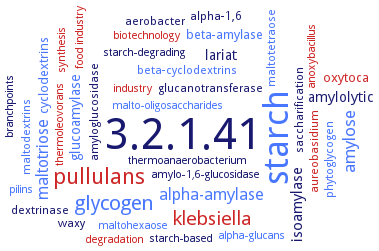
Word Map on EC 3.2.1.41
-
3.2.1.41
-
starch
-
pullulans
-
glycogen
-
klebsiella
-
amylose
-
maltotriose
-
alpha-amylase
-
isoamylase
-
lariat
-
glucoamylase
-
amylolytic
-
beta-amylase
-
oxytoca
-
cyclodextrins
-
dextrinase
-
amyloglucosidase
-
alpha-1,6
-
saccharification
-
beta-cyclodextrins
-
waxy
-
maltodextrins
-
aureobasidium
-
glucanotransferase
-
phytoglycogen
-
maltotetraose
-
aerobacter
-
synthesis
-
pilins
-
thermoanaerobacterium
-
branchpoints
-
starch-based
-
alpha-glucans
-
malto-oligosaccharides
-
starch-degrading
-
maltohexaose
-
anoxybacillus
-
industry
-
food industry
-
degradation
-
biotechnology
-
thermoleovorans
-
amylo-1,6-glucosidase
- 3.2.1.41
- starch
- pullulans
- glycogen
- klebsiella
- amylose
- maltotriose
- alpha-amylase
- isoamylase
-
lariat
- glucoamylase
-
amylolytic
- beta-amylase
- oxytoca
- cyclodextrins
-
dextrinase
- amyloglucosidase
-
alpha-1,6
-
saccharification
- beta-cyclodextrins
-
waxy
- maltodextrins
- aureobasidium
-
glucanotransferase
- phytoglycogen
- maltotetraose
-
aerobacter
- synthesis
- pilins
-
thermoanaerobacterium
-
branchpoints
-
starch-based
- alpha-glucans
- malto-oligosaccharides
-
starch-degrading
- maltohexaose
- anoxybacillus
- industry
- food industry
- degradation
- biotechnology
- thermoleovorans
- amylo-1,6-glucosidase
Reaction
Synonyms
1,4-alpha-D-glucan glucanohydrolase, Alpha-dextrin endo-1,6-alpha-glucosidase, amylase-pullulanase, amylopectin 6-glucanohydrolase, amylopullulanase, AmyX, Apu, ApuK885, ApuM957, Ask, BaPul13A, BDPulA324, BmPul, BNPulA324, CAC60157, debranching enzyme, EC 3.2.1.69, glucanohydrolase, amylopectin 6-, limit dextrinase, More, Nostoc punctiforme debranching enzyme, NPDE, PbPulA, Pcal-1616, PelBsp-PulA, PfAPU, Promozyme 200 L, PUL US105, Pul13A, Pul3YH5, PulA, PulASK, PulB, pulGT, Pullulan 6-glucanohydrolase, pullulan-6-glucanohydrolase, pullulanase, pullulanase type 1, pullulanase type I, pullulanase type II, PulWB42, R-enzyme, Saci_1162, TPApu, TTC1198, TTHpu, type II pullulanase, ZUP1
ECTree

Advanced search results
Crystallization
Crystallization on EC 3.2.1.41 - pullulanase
Please wait a moment until all data is loaded. This message will disappear when all data is loaded.
 top
topCRYSTALLIZATION (Commentary) 

ORGANISM
UNIPROT
LITERATURE
X-ray diffraction structure determination and analysis at 1.7 A resolution
-
hanging dop method, crystals belong to the orthorhombic space group P2(1)2(1)2(1) with unit-cell parameters a = 70.568, b = 127.68, c = 189.25 A. The crystal contains two molecules of pullulanase in the asymmetric unit, with a solvent content of 53.15%
-
hanging-drop vapor diffusion method. Characterization of the Ins subdomain by X-ray crystallography
hanging-drop vapour-diffusion method, crystal structure of pullulanase and its complex with glucose, maltose, isomaltose, maltotriose and maltotetraose, 1.7-1.9 A resolution
-
purified recombinant enzyme, mixing of 0.0012 ml of 14 mg/ml protein in 50 mM Na-phosphate buffer, pH 7.5, with 0.0012 ml reservoir solution containing 20% w/v PEG 8000, 200 mM Tris-HCl buffer, pH 8.5, and 100 mM MgCl2, X-ray diffraction structure determination and analysis at 2.37 A resolution, molecular structure modelling
-
N-terminal truncated form lacking the carbohydrate binding module, but containing the catalytic domain, in its apo form and in complex with maltotetraose, at resolutions of 2.1 and 2.4 A, respectively
-