3.2.1.139: alpha-glucuronidase
This is an abbreviated version!
For detailed information about alpha-glucuronidase, go to the full flat file.
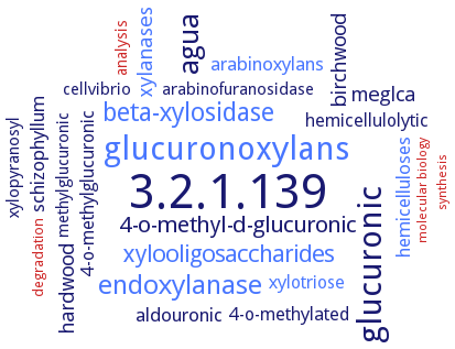
Word Map on EC 3.2.1.139
-
3.2.1.139
-
glucuronoxylans
-
glucuronic
-
agua
-
endoxylanase
-
beta-xylosidase
-
xylooligosaccharides
-
4-o-methyl-d-glucuronic
-
xylanases
-
meglca
-
hardwood
-
birchwood
-
hemicelluloses
-
schizophyllum
-
arabinoxylans
-
aldouronic
-
xylotriose
-
4-o-methylated
-
4-o-methylglucuronic
-
hemicellulolytic
-
methylglucuronic
-
arabinofuranosidase
-
xylopyranosyl
-
cellvibrio
-
analysis
-
degradation
-
synthesis
-
molecular biology
- 3.2.1.139
- glucuronoxylans
-
glucuronic
-
agua
- endoxylanase
- beta-xylosidase
- xylooligosaccharides
-
4-o-methyl-d-glucuronic
- xylanases
-
meglca
-
hardwood
-
birchwood
- hemicelluloses
-
schizophyllum
- arabinoxylans
-
aldouronic
- xylotriose
-
4-o-methylated
-
4-o-methylglucuronic
-
hemicellulolytic
-
methylglucuronic
- arabinofuranosidase
-
xylopyranosyl
-
cellvibrio
- analysis
- degradation
- synthesis
- molecular biology
Reaction
Synonyms
(4-O-methyl)-alpha-glucuronidase, 4-O-methylglucuronidase, aGlu, Agu115, Agu115A, Agu4B, AguA, alpha-(4-O-methyl)-D-glucuronidase, alpha-D-glucuronidase, Alpha-glucosiduronase, alpha-glucosiduronate glucuronohydrolase, alpha-glucuronidase, amylouronate hydrolase-I, Aryl alpha-glucuronidase, AugA, AUH-I, BoAgu115A, DEG75-AG, GH115, GH115 glucuronidase, GH67, GH67 alpha-glucuronidase, GlcA115A, GlcA67A, GLRI, glucuronidase, alpha-, glycosyl hydrolase family 115 alpha-glucuronidase, non-xylanolytic alpha-glucuronidase, p-nitrophenyl alpha-D-glucuronide-hydrolyzing enzyme, Pjdr2_5977, PNP-GAase, RUM630-AG, Sde_1755, TrDCase, TreDCase
ECTree

Advanced search results
Crystallization
Crystallization on EC 3.2.1.139 - alpha-glucuronidase
Please wait a moment until all data is loaded. This message will disappear when all data is loaded.
 top
topCRYSTALLIZATION (Commentary) 

ORGANISM
UNIPROT
LITERATURE
purified recombinant His6-tagged wild-type and selenomethionine-labeled enzyme or enzyme complexed with alpha-D-glucuronic acid, 10 mg/ml protein mixed with 19% PEG3350, 0.2 M sodium citrate, pH 5.5, soaking with 300 mM glucuronic acid for the complexed structure, use of mother liquor supplemented with 15% v/v PEG 400 or paratone N oil as cryoprotectant, X-ray diffraction structure determination and analysis at 2.14-3.0 A resolution
crystal structure of mutant E292A in complex with its substrate aldobiouronic acid
-
hanging drop method, several high resolution crystal structures of the alpha-glucuronidase in complex with its substrate and products: structure of wild-type enzyme, structure of mutant enzyme E285N, mutant enzyme in complex with aldotetraouronic acid
-
hanging-drop vapor diffusion method. Two crystal forms: T1 and M1. T1 form: space group P4(1)2(1)2 or P4(3)2(1)2 with unit-cell dimensions a = b = 76.1 A, c = 331.2 A. The crystals are mechanically strong, are stable in the X-ray beam and diffract X-rays to better than 2.4 A resolution. M1 form: space group P2(1) with unit-cell dimensions a = 65.8, b = 127.4, c = 96.6 A and beta = 97.9°. The crystals are quite strong and stable and diffract to better than 2.8 A resolution
-
molecular modeling of structure. The likely catalytic residues are Asp303 (nucleophile) and Glu350 (proton donor). An arginine (Arg299 in Pjdr2) is conserved in all the Agu115 enzymes considered. The xylose-binding cleft bounded by Trp249 and Val426 in Agu115A is predicted to be lined by Trp272 and Met401 in the Agu115A model
structure reveals a five-domain architecture, with an additional insertion C+ domain that has significant impact on the domain arrangement of the protein monomer and its dimerization. The participation of domain C+ in substrate binding is supported by reduced substrate inhibition upon introducing W773A, W689A, and F696A substitutions within this domain. In addition to Asp335, residue Glu216 is essential for the catalytic activtiy
sitting drop vapor diffusion method, using 20% (w/v) PEG 2000 MME and 0.1 M Tris pH 7.0