3.1.4.53: 3',5'-cyclic-AMP phosphodiesterase
This is an abbreviated version!
For detailed information about 3',5'-cyclic-AMP phosphodiesterase, go to the full flat file.
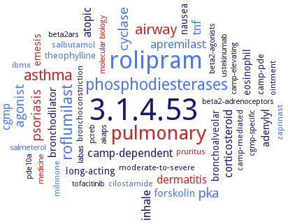
Word Map on EC 3.1.4.53
-
3.1.4.53
-
rolipram
-
pulmonary
-
phosphodiesterases
-
roflumilast
-
asthma
-
airway
-
cyclase
-
pka
-
psoriasis
-
agonist
-
apremilast
-
inhale
-
tnf
-
camp-dependent
-
atopic
-
dermatitis
-
corticosteroid
-
cgmp
-
forskolin
-
bronchodilator
-
adenylyl
-
eosinophil
-
nausea
-
bronchoalveolar
-
emesis
-
long-acting
-
theophylline
-
camp-pde
-
salbutamol
-
ointment
-
ibmx
-
pruritus
-
cilostamide
-
moderate-to-severe
-
milrinone
-
bronchoconstriction
-
camp-mediated
-
zaprinast
-
medicine
-
salmeterol
-
pcreb
-
beta2ars
-
beta2-adrenoceptors
-
labas
-
camp-elevating
-
beta2-agonists
-
pde10a
-
cgmp-specific
-
molecular biology
-
akaps
-
tofacitinib
-
ustekinumab
- 3.1.4.53
- rolipram
- pulmonary
- phosphodiesterases
- roflumilast
- asthma
- airway
- cyclase
- pka
- psoriasis
- agonist
- apremilast
-
inhale
- tnf
-
camp-dependent
-
atopic
- dermatitis
-
corticosteroid
- cgmp
- forskolin
-
bronchodilator
-
adenylyl
-
eosinophil
-
nausea
-
bronchoalveolar
- emesis
-
long-acting
- theophylline
- camp-pde
- salbutamol
-
ointment
- ibmx
- pruritus
- cilostamide
-
moderate-to-severe
- milrinone
-
bronchoconstriction
-
camp-mediated
- zaprinast
- medicine
- salmeterol
-
pcreb
-
beta2ars
-
beta2-adrenoceptors
-
labas
-
camp-elevating
-
beta2-agonists
- pde10a
-
cgmp-specific
- molecular biology
-
akaps
-
tofacitinib
-
ustekinumab
Reaction
Synonyms
3',5'-cAMP phosphodiesterase, 3',5'-cyclic AMP phosphodiesterase, adenosine 3',5'-cyclic monophosphate PDE, adenosine 3',5'-cyclicmonophosphate-specific PDE, cAMP phosphodiesterase, cAMP phosphodiesterase-4, cAMP-PDE, cAMP-phosphodiesterase, cAMP-phosphodiesterase 1C, cAMP-phosphodiesterase 4D7, cAMP-specific 3',5'-cyclic phosphodiesterase 4D, cAMP-specific cyclic nucleotide phosphodiesterase, cAMP-specific PDE, cAMP-specific PDE4A5, cAMP-specific PDE4D2, cAMP-specific phosphodiesterase, cAMP-specific phosphodiesterase 4D, cAMP-specific phosphodiesterase 4D11, cAMP-specific phosphodiesterase-4D5, cAMPspecific PDE, class I phosphodiesterase PDEB1, class I phosphodiesterase PDEB2, CpdA, CPDS, cyclic AMP phosphodiesterase type 4, cyclic AMP phosphodiesterase-4, cyclic AMP-specific phosphodiesterase, cyclic nucleotide phosphodiesterase, cyclic nucleotide phosphodiesterase 4, cyclic nucleotide phosphodiesterase 4D, cyclic nucleotide phosphodiesterase-8A, DdPDE4, high affinity cAMP-specific and IBMX-insensitive 3',5'-cyclic phosphodiesterase 8A, hPDE4A, hPDE4B, HSPDE4A4B, LmjPDEB1, LmjPDEB2, PDE, PDE IVB, PDE-46, PDE-4D3, PDE10A, PDE1C, PDE2, PDE3, PDE4, PDE4A, PDE4A cAMP-specific phosphodiesterase splice variant RD1, PDE4A1, PDE4A10, PDE4A4, PDE4A5, PDE4A8, PDE4B, PDE4B1, PDE4B2, PDE4B3, PDE4B4, PDE4B5, PDE4C, PDE4C2, PDE4D, PDE4D1, PDE4D11, PDE4D3, PDE4D5, PDE4D7, PDE5, PDE7, PDE7A, PDE7A1, PDE7A2, PDE7B, PDE8, PDE8A, PDE8A1, PDE8B, PdeA, PdeB, PDEB1, PdeH, PdeL, phosphodiesterase 4, phosphodiesterase 4 isoform A8, phosphodiesterase 4B, phosphodiesterase 4D, phosphodiesterase 5, phosphodiesterase 7, phosphodiesterase 7B, phosphodiesterase type 4, phosphodiesterase type 5, phosphodiesterase-4, phosphodiesterase-4A5, phosphodiesterase-4B, RegA, RNPDE4A1A, TbPDE1, TbPDE2B, TbrPDEB1, TbrPDEB2, TcPDE1, TcPDE4, TcrPDEA1, TcrPDEB1
ECTree

Advanced search results
Crystallization
Crystallization on EC 3.1.4.53 - 3',5'-cyclic-AMP phosphodiesterase
Please wait a moment until all data is loaded. This message will disappear when all data is loaded.
 top
topCRYSTALLIZATION (Commentary) 

ORGANISM
UNIPROT
LITERATURE
catalytic domain of inactive mutant D201N in complex with substrate cAMP at 1.56 A resolution. Q369 forms only one hydrogen bond ith the adenine of cAMP. Structural comparison between isoform PDE4D2-cAMP and PDE10A2-cAMP shows an anti configuration of cAMP in PDE4, but syn in PDE10
-
hanging drop method, PDE4D2 in complex with the nonselective inhibitor 3-isobutyl-1-methylxanthine
-
hanging-drop vapor-diffusion method, crystal structures of the catalytic domain of phosphodiesterase 4B complexed with AMP (2.0 A), 8-Br-AMP (2.13 A), and rolipram (2.0 A)
-
in complex with inhibitor 4-[8-(3-nitrophenyl)-[1,7]naphthyridin-6-yl]benzoic acid, comparison with isoforms PDE4A, PDE4B, PDE4C. Inhibitor binds in the same conformation to the deep cAMP substrate pocket and interacts with the same residues in each instance. Detailed structural comparison
in complex with inhibitor 4-[8-(3-nitrophenyl)-[1,7]naphthyridin-6-yl]benzoic acid, comparison with isoforms PDE4A, PDE4C, PDE4D. Inhibitor binds in the same conformation to the deep cAMP substrate pocket and interacts with the same resiudues in each instance. Detailed structural comparison
in complex with inhibitor 4-[8-(3-nitrophenyl)-[1,7]naphthyridin-6-yl]benzoic acid, comparison with isoforms PDE4B, PDE4C, PDE4D. Inhibitor binds in the same conformation to the deep cAMP substrate pocket and interacts with the same residues in each instance. Detailed structural comparison
molecular dynamics simulations. The second bridging ligand in the active site is HO- rather than H2O, serving as a nucleophile to initialize the catalytic hydrolysis of cAMP
-
NMR and CD analysis of the N-terminal 38mer peptide of isoform PDE4D5 which contains the entire signaling scaffold protein RACK1 interaction domain together with a portion of the beta-arrestin binding site. The peptiode has a distinct amphipathic helical structure. Study on binding to RACK1 and to beta-arrestin
unliganded PDE8A1 and in complex with 3-isobuytl-1-methylxanthine, hanging drop vapour diffusion method, using 100 mM sodium cacodylate (pH 6.5), 15% 2-propanol, 30% ethylene glycol, and 8-10% PEG3350 at 4°C
unliganded, detailed structural comparison with isoforms PDE4A, PDE4B, PDE4C