the enzyme forms a dimer with each monomer consisting of an N-terminal GAF domain connected to a C-terminal HD-GYP domain by an approximately 42 residue-long helix. Assembly of the head-to-head dimer relies exclusively on the GAF domain and the long alpha5 helix with the HD-GYP domain playing no role in the dimeric interface, crstal structure analysis, overview
the enzyme forms a dimer with each monomer consisting of an N-terminal GAF domain connected to a C-terminal HD-GYP domain by an approximately 42 residue-long helix. Assembly of the head-to-head dimer relies exclusively on the GAF domain and the long alpha5 helix with the HD-GYP domain playing no role in the dimeric interface, crstal structure analysis, overview
2 * 74500 + 2 * 23000, about, sedimentation equilibrium analysis, the dual-functioning diguanylate cyclase/phosphodiesterase enzyme, formed by heme-nitric oxide/oxygen binding protein and cyclic-di-GMP processing enzyme, builds a tetrameric structure
catalytic activity is confined to the C-terminal EAL domain. The GGDEF domain lacks diguanylate cyclase activity but is able to bind GTP which results in activation of the phosphodiresterase activity in the neighbouring EAL domain
MtbDGC contains cysteine pairs Cys94-Cys584, Cys2-Cys479 and Cys429-Cys614, and one unbound Cys406. Structure-function relationship, homology modeling, minimization and model validation, overview
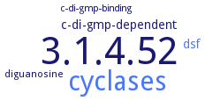
the HD-GYP phosphodiesterase domain of RpfG, which is part of a system necessary for the diffusible signalling factor dependent production of extracellular pathogenicity, interacts directly with diguanylate cyclase GGDEF domain-containing proteins. Physical linkage between quorum-sensing and cyclic diguanylate signalling pathways

 top
top

