1.2.1.19: aminobutyraldehyde dehydrogenase
This is an abbreviated version!
For detailed information about aminobutyraldehyde dehydrogenase, go to the full flat file.
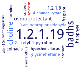
Word Map on EC 1.2.1.19
-
1.2.1.19
-
badhs
-
choline
-
osmoprotectant
-
1.2.1.8
-
spinacia
-
2-acetyl-1-pyrroline
-
glycinebetaine
-
3-aminopropionaldehyde
-
suaeda
-
halophyte
-
fragrant
-
hypochondriacus
-
aldh10s
-
amaranthus
-
liaotungensis
-
jasmine
-
basmati
-
4-aminobutyrate
-
synthesis
- 1.2.1.19
-
badhs
- choline
-
osmoprotectant
-
1.2.1.8
-
spinacia
-
2-acetyl-1-pyrroline
- glycinebetaine
- 3-aminopropionaldehyde
-
suaeda
-
halophyte
-
fragrant
-
hypochondriacus
- aldh10s
-
amaranthus
-
liaotungensis
-
jasmine
-
basmati
- 4-aminobutyrate
- synthesis
Reaction
Synonyms
4-aminobutanal dehydrogenase, 4-aminobutyraldehyde dehydrogenase, ABAL dehydrogenase, ABALDH, ALDH10A8, ALDH10A9, AMADH, AMADH1, AMADH2, aminoaldehyde dehydrogenase, BADH, betaine aldehyde dehydrogenase, dehydrogenase, aminobutyraldehyde, EC 1.5.1.35, gadbh, gamma-aminobutanal dehydrogenase, gamma-aminobutyraldehyde dehydroganase, gamma-aminobutyraldehyde dehydrogenase, gamma-guanidinobutyraldehyde dehydrogenase, MdAMADH1, MdAMADH2, More, NAD+-aminoaldehyde dehydrogenase, NAD+-dependent aminoaldehyde dehydrogenase, PatD, PsAMADH 1, PsAMADH 2, SlAMADH1, SlAMADH2, YdcW, ZmAMADH1a, ZmAMADH1b, ZmAMADH2
ECTree

Advanced search results
Crystallization
Crystallization on EC 1.2.1.19 - aminobutyraldehyde dehydrogenase
Please wait a moment until all data is loaded. This message will disappear when all data is loaded.
 top
topCRYSTALLIZATION (Commentary) 

ORGANISM
UNIPROT
LITERATURE
isozyme AMADH1 in complex with NAD+, hanging drop vapor diffusion method, using 0.1 M HEPES (pH 7.5), 13% (w/v) PEG 6000 and 5% (v/v) 2-methyl-2,4-pentanediol, at 20°C
isozyme AMADH2 in complex with NAD+, hanging drop vapor diffusion method, using 0.1 M HEPES (pH 7.5), 18% (w/v) PEG 4000, 10% (v/v) isopropanol and 0.5% (w/v) n-octyl beta-D-glucopyranoside, at 20°C
wild-type SlAMADH1 with 5 mM NAD+is crystallized over a reservoir containing 23% PEG 1000, 0.1 M HEPES, pH 7.5, mutant E260A of SlAMADH1 with 5 mM NAD+ is crystallized over a reservoir containing 15% PEG 1500, 0.1 M imidazole, pH 7.0, and 10% glycerol, sitting drop vapor diffusion method, cryoprotectant solution consits of mother liquor supplemented with 25% PEG 400 for SlAMADH1 or 20% glycerol for the E260A mutant, X-ray diffraction structure determination and analysis at 1.90 A resolution, molecular replacement method using the dimer structure of PsAMADH2, PDB ID 3IWJ, as a search model
wild-type ZnAMADH1a with 5 mM NAD+ is crystallized over a reservoir containing 16% PEG 4000, 0.1 M HEPES, pH 7.5, and 10% isopropyl alcohol, sitting drop vapor diffusion method, cryoprotection by 20% glycerol in mother liquor, X-ray diffraction structure determination and analysis at 1.95 A resolution, molecular replacement method using the dimer structure of PsAMADH2, PDB ID 3IWJ, as a search model