1.14.14.18: heme oxygenase (biliverdin-producing)
This is an abbreviated version!
For detailed information about heme oxygenase (biliverdin-producing), go to the full flat file.
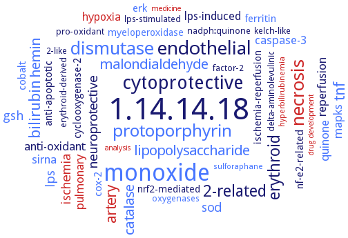
Word Map on EC 1.14.14.18
-
1.14.14.18
-
monoxide
-
cytoprotective
-
endothelial
-
necrosis
-
dismutase
-
protoporphyrin
-
erythroid
-
2-related
-
tnf
-
bilirubin
-
artery
-
malondialdehyde
-
lipopolysaccharide
-
catalase
-
hemin
-
neuroprotective
-
ischemia
-
gsh
-
lps
-
sod
-
anti-oxidant
-
mapks
-
sirna
-
caspase-3
-
quinone
-
pulmonary
-
reperfusion
-
lps-induced
-
hypoxia
-
cyclooxygenase-2
-
cox-2
-
ferritin
-
erk
-
nf-e2-related
-
anti-apoptotic
-
cobalt
-
myeloperoxidase
-
ischemia-reperfusion
-
nrf2-mediated
-
pro-oxidant
-
kelch-like
-
nadph:quinone
-
factor-2
-
lps-stimulated
-
2-like
-
erythroid-derived
-
oxygenases
-
delta-aminolevulinic
-
sulforaphane
-
hyperbilirubinemia
-
medicine
-
drug development
-
analysis
- 1.14.14.18
- monoxide
-
cytoprotective
- endothelial
- necrosis
- dismutase
- protoporphyrin
-
erythroid
-
2-related
- tnf
- bilirubin
- artery
- malondialdehyde
- lipopolysaccharide
- catalase
- hemin
-
neuroprotective
- ischemia
- gsh
- lps
- sod
-
anti-oxidant
- mapks
- sirna
- caspase-3
- quinone
- pulmonary
-
reperfusion
-
lps-induced
- hypoxia
- cyclooxygenase-2
- cox-2
- ferritin
- erk
-
nf-e2-related
-
anti-apoptotic
- cobalt
- myeloperoxidase
-
ischemia-reperfusion
-
nrf2-mediated
-
pro-oxidant
-
kelch-like
-
nadph:quinone
-
factor-2
-
lps-stimulated
-
2-like
-
erythroid-derived
- oxygenases
-
delta-aminolevulinic
- sulforaphane
- hyperbilirubinemia
- medicine
- drug development
- analysis
Reaction
Synonyms
biliverdin-producing heme oxygenase, ChuS, ChuZ, EC 1.14.99.3, haem oxygenase, heme oxygenase, heme oxygenase 1, heme oxygenase 2, heme oxygenase-1, heme oxygenase-2, HemO, Hmox1, Hmox1a, Hmox1b, Hmox2, Hmox2a, Hmox2b, HmuO, Hmx1, HO, HO-1, HO-2, Ho1, Ho2, Ho3, HO4, HSP32, HugZ, HY1, inducible heme oxygenase-1, More, MsHO1, ORP33 proteins, oxygenase, heme (decyclizing), pbsA1, PigA, proteins, specific or class, ORP33 (oxygen-regulated protein 33,000-mol.-wt.), Syn HO-1, Syn HO-2
ECTree

Advanced search results
Crystallization
Crystallization on EC 1.14.14.18 - heme oxygenase (biliverdin-producing)
Please wait a moment until all data is loaded. This message will disappear when all data is loaded.
 top
topCRYSTALLIZATION (Commentary) 

ORGANISM
UNIPROT
LITERATURE
purified recombinant His-tagged enzyme complexed with hemin, hemin:ChuZ ratio of 2:1, microbatch method, mixing of 0..001 ml of 170 mg/ml ChuZ-hemin in 20 mM Tris-HCl, pH 8.0, and 5 mM sodium azide with 0.001 ml of reservoir solution consisting of 0.1 M MES, pH 6.5, 24% PEG400 v/v, and 0.1 M imidazole, X-ray diffraction structure determination and analysis at 2.5 A resolution
-
azide-bound Fe(II)-verdoheme-HmuO, 30°C, a hanging drop vapor diffusion method, the reservoir solution containing 50 mM MES, pH 5.4, 2.3-2.5 M ammonium sulfate, 0.27 M sodium bromide, and 0.1% dioxane, X-ray diffraction structure determination and analysis at 3.0 A resolution
DTT-bound forms of ferric heme-HO-1 complexes, X-ray diffraction structure analysis of crystal structures with PDB IDSs I9T and 3I9U, resolution is 1.5 A
-
hanging drop vapor diffusion method, free enzyme is crystallized by using 85 mM sodium cacodylate, pH 6.5, 25.5% (w/v) PEG8000, 170 mM sodium acetate, and 23.5% (v/v) glycerol, while substrate-bound enzyme is crystallized by using 50 mM MES, pH 6.1, 2.2 M ammonium sulfate, and 25% (w/v) sucrose, supplemented with 200 mM sodium ascorbate
heme oxygenase HmuO in tertiary complex with reaction intermediate verdoheme and N3, X-ray diffraction structure determination and analysis at 1.7 resolution
sitting drop vapour diffusion method using 12-15% (w/v) polyethylene glycol 3350, 0.15-0.20 M magnesium formate, and 10-20 mM NAD
-
purified recombinant HugZ, hanging-drop vapour diffusion method, mixing of 0.001 ml of 25 mg/ml HugZ-hemin in 20 mM Tris, pH 8.0, 5 mM sodium azide, with 0.001 ml of reservoir solution consisting of 16% w/v PEG 3350, 3% Tacsimate, 0.1 M imidazole, pH 7.5, and 12% methanol, X-ray diffraction structure determination and analysis at 1.8-2.3 A resolution
-
apo- and heme-bound truncated HO-2, lacking the three heme regulatory motifs and the membrane binding region, apo-HO-2: hanging drop method, 0.0015 ml of 5 mg/ml protein in 50 mM KCl, 50 mM Tris-HCl, pH 7.5, are mixed with 0.001 ml of well solution containing 40% PEG 1500, 200 mM potassium glutamate, and 100 mM triethanolamine, pH 8.5, at 4°C, heme-bound HO-2: hanging drop vapour diffusion method, 0.0015 ml of 5 mg/ml protein in 50 mM KCl, 50 mM Tris-HCl, pH 7.5, are mixed with 0.001 ml of well solution containing 33% PEG dimethlyether 500, 20 mM MgCl2, and 100 mM HEPES, pH 8.5, at 4°C, X-ray diffraction structure determination and analysis at 2.4 A resolution for the apoenzyme, and at 2.6 A resolution for the heme-bound enzyme
crystal structure determination and analysis of HO-1 in complex with different inhibitors, i.e. 2-[2-(4-chlorophenyl)ethyl]-2-[(1H-imidazol-1-yl) methyl]-1,3-dioxolane, (2R,4S)-2-[2-(4-chlorophenyl)ethyl]-2-[(1H-imidazol-1-yl)methyl]-4-[((5-trifluoromethylpyridin-2-yl)thio)methyl]-1,3-dioxolane, 1-(adamantan-1-yl)-2-(1H-imidazol-1-yl)ethanone, 4-phenyl-1-(1,2,4-1H-triazol-1-yl)butan-2-one, and 1-(1H-imidazol-1-yl)-4,4-diphenyl-2-butanone, overview
heme oxygenase HO1 complexed with reaction intermediate verdoheme, and in tertiary complex with verdoheme and NO, X-ray diffraction structure determination and analysis at 2.2 and 2.1 A resolution, respectively
HO-1 in complex with 1-(adamantan-1-yl)-2-(1H-imidazol-1-yl)ethanone to a resolution of 1.54 A, the coordinating nitrogen atom of His25 is shifted by 0.91 A while the Fe moiety is shifted by 0.85 A. Distal pocket of in the complex is more-open than that of the native holoenzyme. HO-1 in complex with 4-phenyl-1-(1,2,4-1H-triazol-1-yl)butan-2-one to a resolution of 2.20 A, or in complex with (2R,4S)-2-[2-(4-chlorophenyl)ethyl]-2-[(1H-imidazol-1-yl)methyl]-4-[((5-trifluoromethylpyridin-2-yl)thio)methyl]-1,3-dioxolane
HO-1 in complex with 1-(adamantan-1-yl)-2-(1H-imidazol-1-yl)ethanone, by sitting-drop vapor diffusion method, at room temperature, to 1.5 A resolution. Overall structure of the HO-1-inhibitor complex is very similar to that of the native heme-conjugated HO-1, being mostly alpha-helical with the heme sandwiched between the proximal and distal helices. The inhibitor binds to the HO-1 distal pocket such that the imidazolyl moiety coordinates with heme iron while the adamantyl group is stabilized by a hydrophobic binding pocket. Distal helix flexibility, coupled with shifts in proximal residues and heme, acts to expand the distal pocket, thus accommodating the bulky inhibitor without displacing heme. Inhibitor binding effectively displaces the catalytically critical distal water ligand
in complex with ferrous and ferrous-NO forms of verdoheme, absence of network of water-molecules in both structures
mutant H25R with bound heme, to 2.95 A resolution. Contrary to wild-type, the Arg25 side-chain in the H25R mutant is oriented away from the parent His25 position, accompanied by reorientation of the Glu29 side-chain
wild-type and mutant H25A, by hanging drop vapor diffusion method, at 2.8 A resolution. Mutant heme-HO-1 crystal is monoclinic, space group P21, there are four molecules in an asymmetric unit cell. The molecular surfaces of wild-type and mutant are of little difference, though the active pocket in the mutant is larger. The two have the same substrate affinity in electrostatic potential. A positive charge region on the edge of heme forms around the catalytic reaction pocket. After mutation, Ala is still located in the surface and does not influence the electrostatic potential of the reaction pocket
-
heme oxygenase-heme complex at 1.73 A resolution. A hydrogen-bonded network of structural water molecules that extends from the catalytic site to the protein surface is observed. A long-range communication exists between the outer fringes of the hydrogen-bonded network of structural waters and the heme active site during catalysis. Model for the productive association of ferredoxin-NADP+ reductase and heme oxygenase
-
crystal structure at 1.5 A resolution, comparison with heme oxygenase-1 from mammalian sources
-
molecular docking of inhibitor (E)-2-(4-isopropylbenzylidene)hydrazinecarboximidamide to HemO on the back site and the heme site
heme oxygenase HO1 complexed with reaction intermediate verdoheme, X-ray diffraction structure determination and analysis at 2.2 A resolution
HO-1 in complex with (2-[2-(4-chlorophenyl)ethyl]-2-[1H-imidazol-1-yl)methyl]-1,3-dioxolane to 2.70 A resolution, the heme and helix are shifted by ca. 0.8 A toward the alpha-meso carbon along the alpha-gamma axis of the heme
thiol-bound forms of ferric heme-HO-1 complexes, thiols are from DTT or DTE, X-ray diffraction structure analysis of crystal structures with PDB IDSs I9T and 3I9U, resolution is 2.15-2.25 A
truncated heme oxygenase-1, hanging-drop vapor diffusion, enzyme solution is mixed with an equal volume of each reservoir solution and equilibrated, crystals are obtained at 293 K, reservoir solution contains 4 M sodium formate, 18 mg/ml enzyme concentration in 50 mM potassium phosphate, pH 7.0, hexagonal rod-shaped crystals appear after 3 d, X-ray structure of heme oxygenase in complex with heme bound to azide at 1.9 A resolution
truncated, soluble hHO-1 variant, consisting of 233 amino acids, in complex with 4-phenyl-1-(1H-1,2,4-triazol-1-yl)-2-butanone, sitting-drop vapor diffusion, room temperature, 0.412 mM heme-HO-1 complex in 20 mM potassium phosphate is mixed with inhibitor in a 1:3 molar ratio, the reservoir solution contains100 mM HEPES, pH 7.5, 2.00-2.28 M ammonium sulfate, and 0.8-1.1% 1,6-hexanediol, X-ray diffraction structure determination and analysis
-
mutant H25R with bound heme, to 2.95 A resolution. Contrary to wild-type, the Arg25 side-chain in the H25R mutant is oriented away from the parent His25 position, accompanied by reorientation of the Glu29 side-chain