1.1.1.36: acetoacetyl-CoA reductase
This is an abbreviated version!
For detailed information about acetoacetyl-CoA reductase, go to the full flat file.
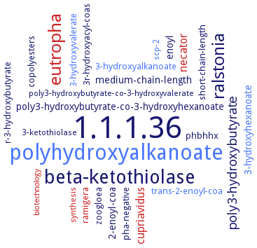
Word Map on EC 1.1.1.36
-
1.1.1.36
-
polyhydroxyalkanoate
-
beta-ketothiolase
-
eutropha
-
ralstonia
-
poly3-hydroxybutyrate
-
cupriavidus
-
necator
-
medium-chain-length
-
2-enoyl-coa
-
3-hydroxyhexanoate
-
enoyl
-
poly3-hydroxybutyrate-co-3-hydroxyhexanoate
-
short-chain-length
-
ramigera
-
3-hydroxyalkanoate
-
3r-hydroxyacyl-coas
-
trans-2-enoyl-coa
-
phbhhx
-
pha-negative
-
zoogloea
-
r-3-hydroxybutyrate
-
copolyesters
-
3-hydroxyvalerate
-
poly3-hydroxybutyrate-co-3-hydroxyvalerate
-
3-ketothiolase
-
scp-2
-
synthesis
-
biotechnology
- 1.1.1.36
- polyhydroxyalkanoate
- beta-ketothiolase
- eutropha
-
ralstonia
-
poly3-hydroxybutyrate
- cupriavidus
- necator
-
medium-chain-length
-
2-enoyl-coa
- 3-hydroxyhexanoate
-
enoyl
-
poly3-hydroxybutyrate-co-3-hydroxyhexanoate
-
short-chain-length
- ramigera
- 3-hydroxyalkanoate
-
3r-hydroxyacyl-coas
- trans-2-enoyl-coa
-
phbhhx
-
pha-negative
-
zoogloea
-
r-3-hydroxybutyrate
-
copolyesters
- 3-hydroxyvalerate
-
poly3-hydroxybutyrate-co-3-hydroxyvalerate
- 3-ketothiolase
- scp-2
- synthesis
- biotechnology
Reaction
Synonyms
(3R)-hydroxyacyl-CoA dehydrogenase, (R)-3-hydroxyacyl-CoA dehydrogenase, AAR, AcAc-CoA reductase, acetoacetyl CoA reductase, acetoacetyl coenzyme A reductase, acetoacetyl-CoA reductase, AKR1B15, aldo-keto reductase 1B15, beta-ketoacyl reductase, beta-ketoacyl-CoA reductase, beta-ketoacyl-coenzyme A reductase, D(-)-beta-hydroxybutyryl CoA-NADP oxidoreductase, D-3-hydroxyacyl-CoA dehydrogenase, D-3-hydroxyacyl-CoA reductase, D-specific 3-hydroxyacyl-CoA dehydrogenase, hydroxyacyl coenzyme-A dehydrogenase, KCR, ketoacyl reductase, MFE-2, More, MSMEG_6753, NADH-preferring acetoacetyl-CoA reductase, NADP-linked acetoacetyl CoA reductase, NADPH-dependent acetoacetyl coenzyme A reductase, NADPH-dependent acetoacetyl-CoA reductase, NADPH-dependent acetoacetyl-coenzyme A reductase, NADPH-linked acetoacetyl-CoA reductase, NADPH:acetoacetyl-CoA reductase, PHA-specific acetoacetyl-CoA reductase, PhaB, PhbB, polyhydroxyalkanoate-specific acetoacetyl coenzyme A reductase, polyhydroxybutyrate enzyme, short chain beta-ketoacetyl(acetoacetyl)-CoA reductase
ECTree

Advanced search results
Crystallization
Crystallization on EC 1.1.1.36 - acetoacetyl-CoA reductase
Please wait a moment until all data is loaded. This message will disappear when all data is loaded.
 top
topCRYSTALLIZATION (Commentary) 

ORGANISM
UNIPROT
LITERATURE
5 mg/ml purified recombinant, detagged truncated enzyme, amino acid residues 1-604, by vapor diffusion technique, 20°C, in 30% PEG 4000, 0.1 M sodium acetate, pH 4.2, 0.2 M ammonium acetate, equal volume of 0.002 ml of protein and precipitant solution, 11% v/v glycerol as cryoprotectant, X-ray diffraction structure determination and analysis at 2.22-2.36 A resolution, the wild-type enzyme, comprising amino acid residues 1-906, is unstable and not crystallizable
-
purified recombinant His-tagged wild-type and mutant enzymes in complex with NADP+ and acetoacetyl-CoA, wild-type protein crystals grow from 0.1 M MES, pH 7.1, 0.9 mM NADP+, 0.9 mM acetoacetyl-CoA, 1.6 M ammonium sulfate, and 10% 1,4-dioxane, mutant crystals grow from 0.6-1.2 M sodium-potassium tartrate, 0.16-0.20 M lithium sulfate, and 0.1 M CHES, pH 8.9-9.9, X-ray diffraction structure determination and analysis at 1.8 A and 2.0-2.9 A resolution, respectively, molecular replacement method using the structure of FabG from Escherichia coli, PDB ID 1I01, as the search probe
apo and NADP+-bound enzyme forms, sitting drop vapor diffusion method, using 100 mM Na-HEPES (pH 7.5), 30% (v/v) PEG 400 and 200 mM magnesium chloride hexahydrate for the apo enzyme and 200 mM sodium citrate tribasic dehydrate and 20% (w/v) PEG 3350 for the NADP+-bound enzyme
selenomethionine-labeled recombinant truncated enzyme, X-ray diffraction structure determination and analysis at 2.38 A resolution
-
sitting drop vapor diffusion method, using 10% (w/v) PEG 8000, 8% (w/v) ethylene glycol, 100 mM HEPES-NaOH pH 7.5