1.1.1.239: 3alpha(17beta)-hydroxysteroid dehydrogenase (NAD+)
This is an abbreviated version!
For detailed information about 3alpha(17beta)-hydroxysteroid dehydrogenase (NAD+), go to the full flat file.
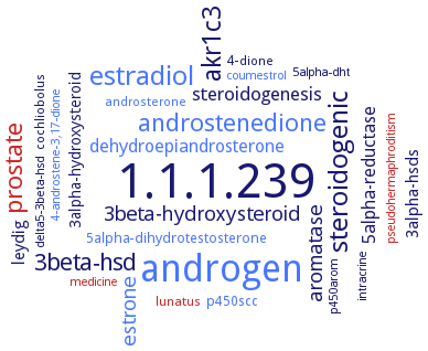
Word Map on EC 1.1.1.239
-
1.1.1.239
-
androgen
-
estradiol
-
steroidogenic
-
akr1c3
-
androstenedione
-
prostate
-
3beta-hsd
-
3beta-hydroxysteroid
-
estrone
-
aromatase
-
steroidogenesis
-
5alpha-reductase
-
dehydroepiandrosterone
-
leydig
-
3alpha-hsds
-
3alpha-hydroxysteroid
-
p450scc
-
5alpha-dihydrotestosterone
-
cochliobolus
-
4-dione
-
androsterone
-
lunatus
-
delta5-3beta-hsd
-
intracrine
-
coumestrol
-
5alpha-dht
-
pseudohermaphroditism
-
p450arom
-
4-androstene-3,17-dione
-
medicine
- 1.1.1.239
- androgen
- estradiol
-
steroidogenic
- akr1c3
- androstenedione
- prostate
- 3beta-hsd
-
3beta-hydroxysteroid
- estrone
- aromatase
-
steroidogenesis
-
5alpha-reductase
- dehydroepiandrosterone
- leydig
- 3alpha-hsds
-
3alpha-hydroxysteroid
- p450scc
- 5alpha-dihydrotestosterone
-
cochliobolus
-
4-dione
- androsterone
- lunatus
- delta5-3beta-hsd
-
intracrine
- coumestrol
-
5alpha-dht
- pseudohermaphroditism
- p450arom
- 4-androstene-3,17-dione
- medicine
Reaction
Synonyms
17-ketoreductase, 17beta-HSD, 17beta-hydroxysteroid dehydrogenase type 5, 3,17beta-HSD, 3,17beta-hydroxysteroid dehydrogenase, 3alpha(17beta)-HSD, 3alpha(17beta)-hydroxysteroid dehydrogenase, AKR1C26, AKR1C27, AKR1C28, AKR1C3, AKR1C34, aldo-keto reductase 1C3, dehydrogenase, 3alpha,17beta-hydroxy steroid, EC 1.1.1.63, More, NAD+-dependent 3alpha(17beta)-hydroxysteroid dehydrogenase, NAD+-dependent 3beta(17beta)-hydroxysteroid dehydrogenase, NAD+-linked S-tetralol dehydrogenase, PGER6, type 2 3alpha-HSD, type 2 3alpha-hxdroxysteroid dehydrogenase/type 5 17beta-hydroxysteroid dehydrogenase, type 2 3alppha-hydroxysteroid dehydrogenase/type 5 17beta-hydroxysteroid dehydrogenase, type 3 17beta-hydroxysteroid dehydrogenase, type 5 17beta-HSD, type 5 17beta-hydroxysteroid dehydrogenase, type 5 17beta-hydroxysteroid dehydrogenase/prostaglandin F synthase, type 5 beta-hydroxysteroid dehydrogenase
ECTree

Advanced search results
Application
Application on EC 1.1.1.239 - 3alpha(17beta)-hydroxysteroid dehydrogenase (NAD+)
 top
top

